-
-
- Total knee replacement: both sides of the joint are replaced by metal or plastic implants.
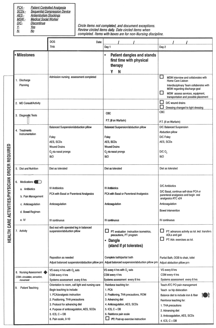
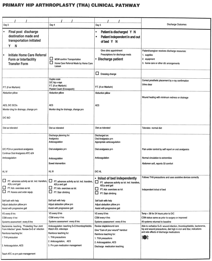
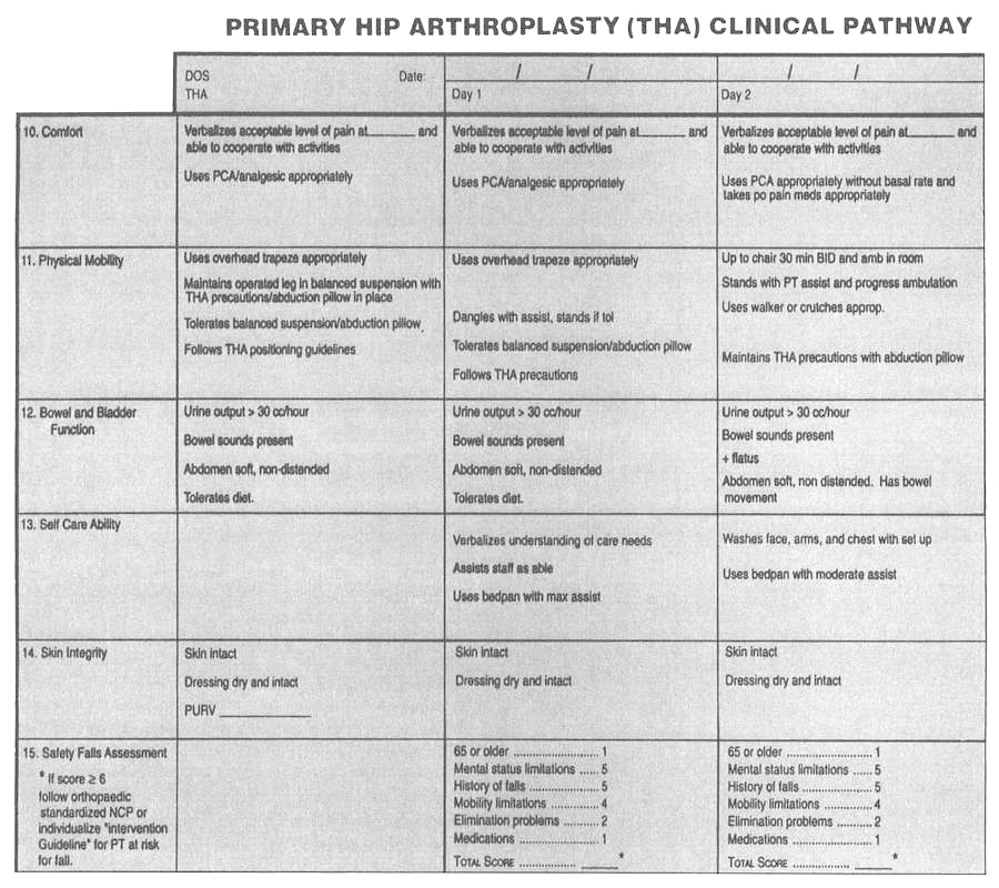
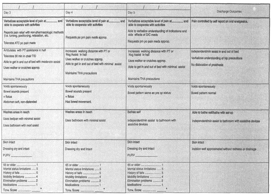
- Analysis: Total hip replacement
- Nursing plan/implementation:
- Goal:
achieve active flexion beyond 70 degrees.
- Immediately postop:
may have continuous passive motion (CPM) device for flexion/extension of affected kne Maximum flexion 110 degrees.
- Monitor drainage in
Hemovac
(q15 minutes for first 4 hours, q1h until 24 hours; q4h and prn while Hemovac in place).
- Analgesics as ordered for pain.
- While dressings are still on: quadricepssetting exercises for approximately 5 days (consult with physical therapist for specific instructions).
- After dressings removed: active flexion exercises.
- Avoid
pressure on heel.
- Evaluation/outcome criteria:
- No complications of infection, hemorrhage note
- ROM of knee increases with exercises.
- Amputation: surgical removal of a limb as a result of trauma or circulatory impairment (gangrene). The amount of tissue amputated is determined by the severity of disease or trauma and the ability of the remaining tissue to heal.
- Risk factors:
- Atherosclerosis obliterans.
- Uncontrolled diabetes mellitus.
- Malignancy.
- Extensive and intractable infection.
- Result of severe trauma.
- Assessment
(preoperative):
- Subjective data:
pain in affected part.
- Objective data:
- Soft-tissue damag
- Partial or complete severance of a body part.
- Lack of peripheral pulses.
- Skin color changes, pallor —> cyanosis —> gangren
- Infection, hemorrhage, or shock.
- Analysis/nursing diagnosis:
- Impaired physical mobility
related to lowerlimb amputation.
- Body image disturbance
related to loss of body part.
- Pain
related to interruption of nerve pathways.
- Anxiety
related to potential change in lifestyl
- Knowledge deficit
related to rehabilitation goals.
- Nursing care plan/implementation:
- Goal:
prepare for surgery, physically and emotionally.
- Validate that patient and family are aware that amputation of body part is planne
- Validate that informed consent is signed, Allow time for grievin
- Allow time for grievin
- If time allows, prepare patient for postoperative phase, , teach arm-strengthening exercises if lower limb is to be amputated; teach alternative methods of ambulation.
- Provide time to discuss feelings.
- Prepare surgical site to decrease possibility of infection (, shave, scrub as ordered).
- Discuss postoperative expectations.
- Goal:
promote healing postoperatively.
- Monitor respiratory status ql-4h and prn: rate, depth of respiration; auscultate for signs of congestion; and question patient about chest pain (pulmonary emboli common complication).
- Monitor for hemorrhage; keep tourniquet at bedsid
- Medicate for pain as ordered-patient may have phantom limb pain.
- Support stump on pillow for first 24 hours; remove pillow after 24 hours to prevent contractur
- Position: turn patient on to stomach
to prevent hip contractur
- ROM exercises for joint above amputation to prevent joint immobilization; strengthening exercises for arms, nonaffected limbs, abdominal muscles.
- Stump care:
- Early postoperative dressings changed prn.
- As incision heals, bandage is applied in cone shape to prepare stump for prosthesis.
- Inspect for blisters, redness, abrasions.
- Remove stump sock daily and prn.
- Assist in rehabilitation program.
- Evaluation/outcome criteria:
- Begins rehabilitation program.
- No hemorrhage, infection.
- Adjusts to altered body imag
- Gout: disorder of purine metabolism; genetic disease believed to be transmitted by a dominant gene, characterized by recurrent attacks of acute pain and swelling of one joint (usually the great toe).
- Pathophysiology: urate crystals and infiltrating leukocytes appear to damage the intracellular phagolysosomes, resulting in leakage of lysomal enzymes into the synovial fluid, causing tissue damage and joint inflammation.
- Risk factors:
- Men.
- Age (>50).
- Genetic/familial tendency.
- Prolonged hyperuricemia (elevated serum uric acid).
- Obesity.
- Moderate to heavy alcohol intak
- Hypertension.
- Abnormal kidney function.
- Assessment:
- Subjective data:
- Pain: excruciating.
- Fatigue.
- Anorexia.
- Objective data:
- Joint: erythema (redness), hot, swollen, difficult to move; skin stretched and shiny over joint.
- Subcutaneous nodules, tophi (deposits of urate) on hands and feet.
- Weight loss.
- Fever.
- Sensory changes, with cold intoleranc
- Lab data:
- Serum uric acid: increased significantly (6.5/100 mL in women, 7.5/100 mL in men) in chronic gout; only slightly increased in acute gout.
- WBC: 12,000-15,000/μL.
- Erythrocyte sedimentation rate: >20 mm.
- 24 hours urinary uric acid: slightly elevate
- Proteinuria (chronic gout).
- Azotemia (presence of nitrogencontaining compounds in blood) in chronic gout.
- Diagnostic tests: arthrocentesis, x-rays.
- Analysis/nursing diagnosis:
- Pain related to inflammation and swelling of affected joint.
- Impaired physical mobility related to pain.
- Knowledge deficit related to diet restrictions and increased fluid needs.
- Altered urinary elimination related to kidney damage.
- Nursing care plan/implementation:
- Goal: decrease discomfort.
- Administer antigout medications as ordered:
- Treatment of acute attacks: colchicine, phenylbutazone (Butazolidin), indomethacin (Indocin), allopurinol (Zyloprim), naproxen (Naprosyn), corticosteroids (prednisone).
- Preventive therapy: probenecid (Benemid), sulfinpyrazone (Anturane). These drugs are not used during acute attacks.
- Absolute rest of affected joint-gradual increase in activities, to prevent complications of immobilization; at the same time, rest for comfort.
- Goal: prevent kidney damag
- Increase fluid intake to 2000-3000 mL/day.
- Monitor urinary output.
- Goal: health teachin
- Need for low-purine diet during acute attack.
- Importance of increased fluid in diet.
- Signs and symptoms of increased progression of diseas
- Dosage and side effects of prescribed medications.
- Evaluation/outcome criteria:
- Swelling decrease
- Discomfort alleviate
- Mobility returned to status before attack.
- Lab values return to normal.