1. Traumatic injuries to the brain
    1. Primary trauma:
      1. Concussion- transient disorder due to injury in which there is brief loss of consciousness due to paralysis of neuronal function; recovery is usually total.
      2. Contusion- structural alteration of brain tissue characterized by extravasation of blood cells (bruising); injury may occur on side of impact or on opposite side (when cranial contents shift forcibly within the skull with impact).
      3. Laceration- tearing of brain tissue or blood vessels due to a sharp bone fragment or object or tearing force.
      4. Fracture- linear (may result in epidural bleed); comminuted or depressed (may tear dura and result in CSF leak); basilar (most serious). Basilar skull fracture may result in meningitis or brain abscess; bleeding from nose or ears; CSF present in drainage; bruising over mastoid process (Battles sign) and periorbital ecchymosis (raccoon eyes).
    2. Secondary trauma (response to primary trauma):
      1. Hematomas:
        1. Subdural- blood from ruptured or torn vein collects between arachnoid and dura; may be acute, subacute, or chronic.
        2. Extradural (epidural)-blood clot located between dura mater and inner surface of skull; most often from tearing of middle meningeal artery; emergency condition.
      2. Increased intracranial pressure
    3. Mechanisms of injury:
      1. Deformation (blow to the head).
      2. Acceleration-deceleration (coup-contrecoup)-forward and rebounding motion.
      3. Rotation (tension, stretching, shearing force).
    4. Pathophysiology of impaired CNS functioning:
      1. Depressed neuronal activity in reticular activating system-depressed consciousness
      2. Depressed neuronal functioning in lower brain stem and spinal cord-depression of reflex activity-decreased eye movements, unequal pupils-decreased response to light stimuli-widely dilated and fixed pupils.
      3. Depression of respiratory center-altered respiratory pattern-decreased rate respiratory arrest.
    5. Risk factors: accidents-automobile, industrial and home, motorcycle, military.
    6. Assessment:
      1. Objective data:
        1. Headache.
        2. Dizziness, loss of balance.
        3. Double vision.
        4. Nausea.
      2. Objective data:
        1. Laceration or abrasion around face or head; profuse bleeding from scalp (highly vascular, poor vasoconstrictive abilities).
        2. Drainage from ears or nose (serosanguineous).
        3. Projectile vomiting, hematemesis.
        4. Vital signs indicating increased intracranial pressure
        5. Neurological exam:
          1. Altered level of consciousness; a numerical assessment, such as the Glasgow Coma Scale, may be used. The lower the score, the poorer the prognosis, generally.
          2. Pupils-equal, round, react to light; or unequal, dilated, unresponsive to light.
          3. Extremities-paresis or paralysis.
          4. Reflexes-hypotonia or hypertonia; Babinski present (flaring of great toe when sole is stroked).
    7. Analysis/nursing diagnosis:
      1. Altered thought processes related to brain trauma.
      2. Sensory/perceptual alteration related to depressed neuronal activity.
      3. Risk for injury related to impaired CNS functioning.
      4. Risk for aspiration related to respiratory depression.
      5. Self-care deficit related to altered level of consciousness.
      6. Ineffective breathing pattern related to CNS trauma.
    8. Nursing care plan/implementation:
      1. Goal: sustain vital functions and minimize or prevent complications.
        1. Patent airway: endotracheal tube or tracheostomy may be ordered.
        2. Oxygen: as ordered (hypoxia increases cerebral edema).
        3. Be Position: semiprone or prone (coma position) with head level to prevent aspiration (keep off back); turn side to side to prevent stasis in lungs.
        4. Vital signs as ordered.
        5. Neurological check: pupils, level of consciousness, muscle strength; report changes.
        6. Seizure precautions: padded side rails.
        7. Medications as ordered:
          1. Steroids (dexamethasone [Decadron]).
          2. Anticonvulsants (phenytoin [Dilantin], phenobarbital).
          3. Analgesics (morphine contraindicated).
        8. Cooling measures or hypothermia to reduce elevated temperature.
        9. Assist with diagnostic tests:
          1. Lumbar puncture (contraindicated with increased intracranial pressure).
          2. Electroencephalogram (EEG).
        10. Diet: NPO for 24 hours, progressing to clear liquids if awake.
        11. Fluids: IVs; nasogastric tube feedings; I&O.
        12. Monitor blood chemistries: sodium imbalance common with head injuries.
      2. Goal: provide emotional support and use comfort measures.
        1. Comfort: skin care, oral hygiene; sheepskins; wrinkle-free linen.
        2. Eyes: lubricate q4h with artificial tears if periocular edema present.
        3. ROM-passive, active; physical therapy as tolerated.
        4. Avoid restraints.
        5. Encourage verbalization of concerns about changes in body image, limitations.
        6. Encourage family communication.
    9. Evaluation/outcome criteria:
      1. Alert, oriented-no residual effects (cognitive processes intact).
      2. No signs of increased intracranial pressure (decreased respirations, increased systolic pressure with widening pulse pressure, bradycardia).
      3. No paralysisregains motor/sensory function.
      4. Resumes self-care activities.

Levels of Consciousness

Stage Characteristics

Alertness


Aware of time and place


Automatism


Aware of time and place but demonstrates abnormality of mood (euphoria to irritability)


Confusion


Inability to think and speak in coherent manner; responds to verbal requests but is unaware of time and place


Delirium


Restlessness and violent activity; may not comply with verbal instructions


Stupor


Quiet and uncommunicative; may appear conscious-sits or lies with glazed look; unable to respond to verbal instructions; bladder and rectal incontinence may occur


Semicoma


Unresponsive to verbal instructions but responds to vigorous or painful stimuli


Coma


Unresponsive to vigorous or painful stimuli


Glasgow Coma Scale

Best eye-opening response

Purposeful and spontaneous:

  • To voice
  • To pain

No response

Untestable

Best verbal response

  • Oriented
  • Disoriented
  • Inappropriate words
  • Incomprehensible sounds
  • Untestable

Best motor response

  • Obeys commands
  • Localizes pain
  • Withdraws to pain
  • Extension to pain
  • No response
  • Untestable
  1. Increased intracranial pressure (ICP): intracranial hypertension associated with altered states of consciousness.
    1. Pathophysiology: increases in intracranial blood volume, cerebrospinal fluid, or brain tissue mass-increased intracranial pressure-impaired neural impulse transmission cellular anoxia, atrophy.
    2. Risk factors:
      1. Congenital anomalies (hydrocephalus).
      2. Space-occupying lesions (abscesses or tumors).
      3. Trauma (hematomas or skull fractures).
      4. Circulatory problems (aneurysms, emboli).
      5. Inflammation (meningitis, encephalitis).
    3. Assessment:
      1. Subjective data:
        1. Headache (early, but nonspecific symptom).
        2. Nausea.
        3. Visual disturbance (diplopia).
      2. Objective data:
        1. Changes in level of consciousness (early sign).
        2. Pupillary changes-unequal, dilated, and unresponsive to light (late sign).
        3. Vital signs-changes are variable.
          1. Blood pressure-gradual or rapid elevation, widened pulse pressure.
          2. Pulse-bradycardia, tachycardia; significant sign is slowing of pulse as blood pressure rises.
          3. Respirations-pattern changes(Cheyne-Stokes, apneusis, Biot’s), deep and sonorous; hiccups.
          4. Temperature-moderate elevation.
        4. Projectile vomiting (more common in children).
        5. Diagnostic test: Head CT-structural changes.
    4. Analysis/nursing diagnosis:
      1. Altered cerebral tissue perfusion related to increased intracranial pressure.
      2. Altered thought processes related to cerebral anoxia.
      3. Ineffective breathing pattern related to compression of respiratory center.
      4. Risk for aspiration related to unconsciousness.
      5. Self-care deficit related to altered level of consciousness.
      6. Impaired physical mobility related to abnormal motor responses.
    5. Nursing care plan/implementation:promote adequate oxygenation and limit further impairment.
      1. Vital signs: report changes immediately.
      2. Patent airway; keep alkalotic, to prevent increased intracranial pressure from elevated CO2; hyperventilate if necessary.
      3. Give medications as ordered:
        1. Hyperosmolar diuretics (mannitol, urea) to reduce brain swelling.
        2. Steroids (dexamethasone [Decadron]) for anti-inflammatory action.
        3. Antacids or H2 antagonist to prevent stress ulcer.
      4. B Position: head of bed elevated 30 degrees.
      5. Fluids: restrict; strict I&O.
      6. Cooling measures to reduce temperature, because fever increases ICP.
      7. Prepare for surgical intervention.
    6. Evaluation/outcome criteria:
      1. No irreversible brain damage-regains consciousness.
      2. Resumes self-care activities.

  2. Craniotomy: excision of a part of the skull (burr hole to several centimeters) for exploratory purpose and biopsy; to remove neoplasms, evacuate hematomas or excess fluid, control hemorrhage, repair skull fractures, remove scar tissue, repair or excise aneurysms, and drain abscesses; produces minimal neurological deficit.
    1. Analysis/nursing diagnosis:
      1. Altered cerebral tissue perfusion related to edema.
      2. Altered thought processes related to disorientation.
      3. Self-care deficit related to continued neurological impairment.
    2. Nursing care plan/implementation:
      1. Preoperative:
        1. Goal: obtain baseline measures.
          1. Vital signs.
          2. Level of consciousness.
          3. Mental, emotional status.
          4. Pupillary reactions.
          5. Motor strength and functioning.
        2. Goal: provide psychological support: listen; give accurate, brief explanations.
        3. Goal: prepare for surgery.
          1. Cut hair; shave scalp (usually done in surgery); save hair if patient/family desire.
          2. Cover scalp with clean towel.
          3. Insert indwelling Foley catheter as ordered.
      2. Postoperative:
        1. Goal: prevent complications and limit further impairment.
          1. Vital signs (indications of complications):
            1. Decreased blood pressure-shock.
            2. Widened pulse pressure-increased ICP.
            3. Respiratory failure-compression of medullary respiratory centers.
            4. Hyperthermia-disturbance of heat-regulating mechanism; infection.
          2. Neurological:
            1. Pupils-ipsilateral dilation (increased ICP), visual disturbances.
            2. Altered level of consciousness.
            3. Altered cognitive or emotional status-disorientation common.
            4. Motor function and strength- hypertonia, hypotonia, seizures.
          3. Blood gases, to monitor adequacy of ventilation.
          4. Dressings: check frequently; aseptic technique; reinforce as necessary.
          5. Observe for:
            1. CSF leakage (glucose-positive drainage from nose, mouth, ears)-report immediately.
            2. Periorbital edema-apply light ice compresses as necessary remove crusts from eyelids.
          6. Check integrity of seventh cranial nerve (facial)-incomplete closure of eyelids.
          7. Position:
            1. Supratentorial surgery (cerebrum)-semi-Fowler’s (30-degree elevation); may not lie on operative side.
            2. Infratentorial (brain stem, cerebellum)-flat in bed (prone); may turn to either side but not onto back.
          8. Fluids: NPO until bowel sounds return, alert, and intact gag, swallow, and cough reflexes present.
          9. Medications as ordered:
            1. Osmotic diuretics (mannitol).
            2. Corticosteroids (dexamethasone [Decadron]).
            3. Mild analgesics (do not mask neurological or respiratory depression).
            4. Stool softeners to prevent constipation and straining.
          10. Orient frequently to person, time, place-to reduce restlessness, confusion.
          11. Side rails up for safety.
          12. Avoid restraints (may increase agitation and ICP).
          13. Ice bags to head to reduce headache.
          14. Activity: assist with ambulation.
        2. Goal: provide optimal supportive care.
          1. Cover scalp once dressings are removed (scarves, wigs).
          2. Deal realistically with neurological deficits-facilitate acceptance, adjustment, independence.
        3. Goal: health teaching.
          1. Prepare for physical, occupational, or speech therapy, as needed.
          2. Activities of daily living.
    3. Evaluation/outcome criteria:
      1. Regains consciousness-is alert, oriented.
      2. Resumes self-care activities within limits of neurological deficits.
  1. Epilepsy: seizure disorder characterized by sudden transient aberration of brain function; associated with motor, sensory, autonomic, or psychic disturbances.
    1. Seizure: involuntary muscular contraction and disturbances of consciousness from abnormal electrical activity.
      1. Brain injury.
      2. Infection (meningitis, encephalitis).
      3. Water and electrolyte disturbances.
      4. Hypoglycemia.
      5. Tumors.
      6. Vascular disorders (hypoxia or hypocapnia).
    2. Risk factors:
    3. Generalized seizures:
      1. Tonic-clonic (grand mal) seizures:
        1. Pathophysiology: increased excitability of a neuron-possible activation of adjacent neurons-synchronous discharge of impulses-vigorous involuntary sustained muscle spasms (tonic contractions). Onset of neuronal fatigue- intermittent muscle spasms (clonic contractions)-cessation of muscle spasms-fatigue.
        2. Assessment:
          1. Subjective data- aura: flash of light; peculiar smell, sound; feelings of fear; euphoria.
          2. Objective data:
            1. Convulsive stage- tonic and clonic muscle spasms, loss of consciousness, breath holding, frothing at mouth, biting of tongue, urinary or fecal incontinence; lasts 2-5 minutes.
            2. Postconvulsion- headache, fatigue (postictal sleep), malaise, vomiting, sore muscles, choking on secretions, aspiration.
      2. Absence (petit mal) seizures:
        1. Pathophysiology: unknown etiology, momentary loss of consciousness (10-20 seconds); usually no recollection of seizure; resumes previously performed action.
        2. Assessment- objective data:
          1. Fixation of gaze; blank facial expression.
          2. Flickering of eyelids.
          3. Jerking of facial muscle or arm.
      3. Minor motor seizures:
        1. Myoclonic -involuntary “lightning-like” jerking contraction of major muscles; may throw person to the floor; no loss of consciousness.
        2. Atonic -brief, total loss of muscle tone; person falls to the floor; loss of consciousness (common in children).
    4. Partial (focal) seizures:
      1. Partial motor: arises from region in motor cortex (posterior frontal lobe); most commonly begins in upper extremities, spreading to face and lower extremity (jacksonian march); noting progression is important in identifying area of cortex involved.
      2. Partial sensory: sensory symptoms occur with partial seizure activity; varies with region in brain; transient.
      3. Partial complex (psychomotor): arises out of anterior temporal lobe; frequently begins with an aura; characteristic feature is automatism (lip smacking, chewing, patting body, picking at clothes); lasts from 2-3 minutes to 15 minutes; do not restrain.
    5. Analysis/nursing diagnosis:
      1. Risk for injury related to convulsive disorder.
      2. Anxiety related to sudden loss of consciousness.
      3. Self-esteem disturbance related to chronic illness.
      4. Impaired social interaction related to selfconsciousness.
    6. Nursing care plan/implementation (generalized seizures):
      1. Goal: prevent injury during seizure.
        1. Do not force jaws open during convulsion.
        2. Do not restrict limbs-protect from injury; place something soft under head (towel, jacket, hands).
        3. Loosen constrictive clothing.
        4. Note time, level of consciousness, type and duration of seizure.
      2. Goal: postseizure care:
        1. Turn on side to drain saliva and facilitate breathing.
        2. Suction as necessary.
        3. Orient to time and place.
        4. Oral hygiene if tongue or cheek injured.
        5. Check vital signs, pupils, level of consciousness.
      3. Goal: prevent or reduce recurrences of seizure activity.
        1. Encourage patient to identify precipitating factors.
        2. Moderation in diet and exercise.
        3. Medications as ordered: phenytoin (Dilantin); phenobarbital; carbamazepine (Tegretol); primidone (Mysoline); valproate (Depacon).
      4. Goal: health teaching.
        1. Medications:
          1. Actions, side effects (apathy, ataxia, hyperplasia of gums).
          2. Complications with sudden withdrawal (status epilepticus -continuous seizure activity; give diazepam per order, O 2).
        2. Attitude toward life and treatment; adhere to medication program.
        3. Clarify misconceptions, fears-especially about insanity, bad genes.
        4. Maintain activities, interests-expect no driving until seizure free for period of time specified by state Department of Motor Vehicles.
        5. Avoid: stress; lack of sleep; emotional upset; alcohol.
        6. Relaxation techniques; stress-management techniques.
        7. Use Medic Alert band or tag.
        8. Appropriate community resources.
    7. Evaluation/outcome criteria:
      1. Avoids precipitating stimuli-achieves seizure control.
      2. Complies with medication regimen.
      3. Retains independence.

  2. Transient Ischemic Attacks (TIAs): temporary, complete, or relatively complete cessation of cerebral blood flow to a localized area of brain, producing symptoms (2-30 minutes) ranging from weakness (“drop attacks”) and numbness to monocular blindness; an important precursor to strokSurgical intervention includes carotid endarterectomy; most common postoperative cranial nerve damage causes vocal cord paralysis or difficulty managing saliva and tongue deviation (cranial nerves VII, X, XI, XII); usually temporary; stroke may also occur.
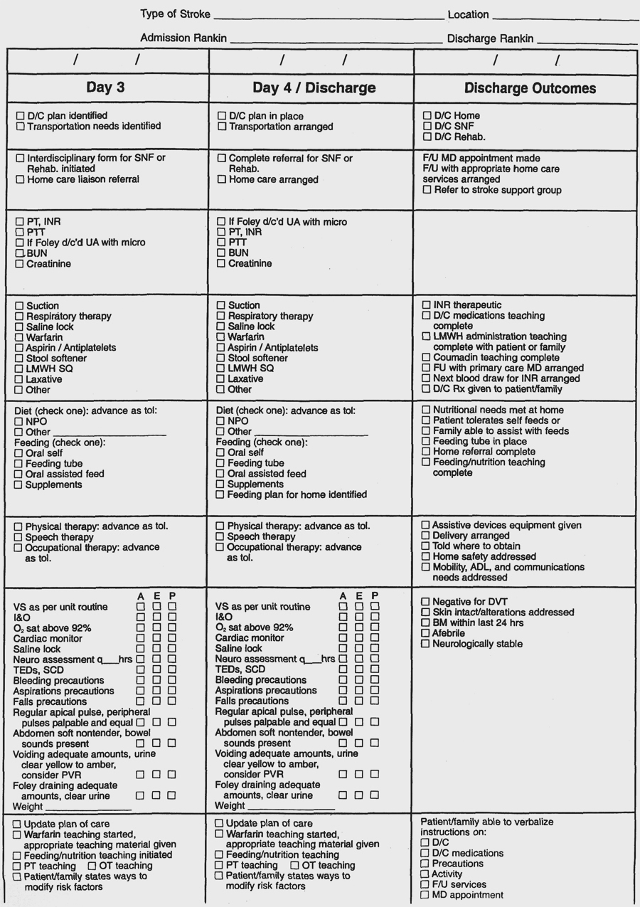
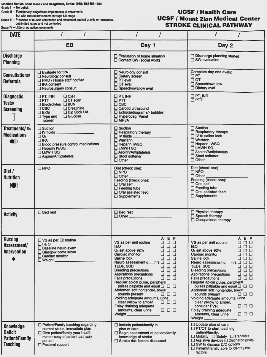
  1. Ischemic stroke (CVA, brain attack): brain lesion resulting from damage to blood vessels supplying brain (middle cerebral artery most common).
    1. Pathophysiology: reduced or interrupted blood flow-interruption of nerve impulses down corticospinal tract-decreased or absent voluntary movement on one side of the body (fine movements are more affected than coarse movements); later, autonomous reflex activity-spasticity and rigidity of muscles.
    2. Risk factors:
      1. Cerebral thrombosis (most common), embolism, hemorrhage.
      2. Prior ischemic episodes (TIAs).
      3. Hypertension.
      4. Oral contraceptives.
      5. Emotional stress.
      6. Family history.
      7. Age.
      8. Diabetes mellitus.
    3. Assessment:
      1. Subjective data:
        1. Weakness: sudden or gradual loss of movement of extremities on one side.
        2. Difficulty forming words.
        3. Difficulty swallowing (dysphagia).
        4. Nausea, vomiting.
        5. History of TIAs.
      2. Objective data:
        1. Vital signs:
          1. BP-elevated; widened pulse pressure.
          2. Temperature-elevated.
          3. Pulse-normal, slow.
          4. Respirations-tachypnea, altered pattern; deep; sonorous.
        2. Neurological:
          1. Altered level of consciousness.
          2. Pupils-unequal; vision-homonymous hemianopia.
          3. Ptosis of eyelid, drooping mouth.
          4. Paresis or paralysis (hemiplegia).
          5. Loss of sensation and reflexes.
          6. Incontinence of urine or feces.
          7. Aphasia
    4. Analysis/nursing diagnosis:
      1. Impaired physical mobility related to hemiplegia.
      2. Impaired swallowing related to paralysis.
      3. Impaired verbal communication related to aphasia.
      4. Risk for aspiration related to unconsciousness.
      5. Sensory/perceptual alterations related to altered cerebral blood flow, visual field blindness.
      6. Altered thought processes related to cerebral edema.
      7. Self-care deficit related to paresis or paralysis.
      8. Body image disturbance related to hemiplegia.
      9. Total incontinence related to interruption of normal nerve transmission.
      10. Impaired social interaction related to aphasia or neurological deficit.
      11. Risk for impaired skin integrity related to immobility.
      12. Unilateral neglect related to cerebral damage
    5. Nursing care plan/implementation:
      1. Goal: reduce cerebral anoxia.
        1. Patent airway:
          1. Oxygen therapy as ordered; suctioning to prevent aspiration.
          2. Turn, cough, deep breathe q2h due to high incidence of aspiration pneumonia.
        2. Activity: bed rest, progressing to out of bed as tolerated.
        3. Position:
          1. Maximize ventilation.
          2. Support with pillows when on side; use hand rolls and arm slings as ordered.
      2. Goal: promote cardiovascular function and maintain cerebral perfusion.
        1. Vital signs; neurological checks.
        2. Medications as ordered:
          1. Antiplatelet aggregation inhibitors (ASA, ticlopidine, dipyridamole [Persantine]).
          2. Antihypertensives to prevent rupture.
          3. Anticoagulants to prevent thrombus.
        3. Fluids: IVs to prevent hemoconcentration; I&O; weigh daily.
        4. ROM exercises to prevent contractures, muscle atrophy, phlebitis.
        5. Skin care and position changes to prevent decubiti.
      3. Goal: provide for emotional relaxation.
        1. Identify grief reaction to changes in body image.
        2. Encourage expression of feelings, concerns.
      4. Goal: patient safety.
        1. Identify existence of homonymous hemianopia (visual field blindness) and agnosia (disturbance in sensory information).
        2. Use side rails and assist as needed.
        3. Remind to walk slowly, take adequate rest periods, ensure good lighting, look where patient is going.
      5. Goal: health teaching.
        1. Exercise routines.
        2. Diet: self-feeding, but assist as needed.
        3. Resumption of self-care activities.
        4. Use of supportive devices; transfer techniques.
    6. Evaluation/outcome criteria:
      1. No complications (pneumonia).
      2. Regains functional independence-resumes self-care activities.
      3. Return of control over body functions (bowel, bladder, speech).


  1. Bacterial meningitis.

  2. Encephalitis (also includes aseptic meningitis): inflammation of the brain and its coverings due to direct viral invasion, which usually results in a lengthy coma.
    1. Pathophysiology: brain tissue injury- release of enzymes that increase vascular dilation, capillary permeability-edema formation-increased intracranial pressure-depression of CNS function.
    2. Risk factors:
      1. Arboviruses.
      2. Enteroviruses (poliovirus, echovirus).
      3. Herpesvirus.
      4. Varicella-zoster (chickenpox).
      5. Postinfection complication (measles, mumps, smallpox).
    3. Assessment:
      1. Subjective data:
        1. Headache-severe.
        2. Fever-sudden.
        3. Nausea, vomiting.
        4. Sensitivity to light (photophobia).
        5. Difficulty concentrating.
      2. Objective data:
        1. Altered level of consciousness.
        2. Nuchal rigidity.
        3. Tremors; facial weakness.
        4. Nystagmus.
        5. Elevated temperature.
        6. Diagnostic test: lumbar puncture-fluid cloudy; increased neutrophils, protein.
        7. Lab data: blood-slight to moderate leukocytosis (about 14,000).
    4. Analysis/nursing diagnosis:
      1. Self-care deficit related to altered level of consciousness.
      2. Risk for injury related to coma.
      3. Sensory/perceptual alteration related to brain tissue injury.
      4. Altered thought processes related to increased intracranial pressure.
    5. Nursing care plan/implementation:
      1. Goal: support physical and emotional relaxation.
        1. Vital signs; neurological signs as ordered.
        2. Seizure precautions.
        3. Position: to maintain patent airway; prevent contractures; ROM.
        4. Medications as ordered:
          1. Analgesics for headache and neck pain.
          2. Antipyretics for fever.
          3. Antivirals.
          4. Anticonvulsants for seizures.
          5. Antibiotics for infection in aseptic meningitis.
          6. Osmotic diuretics to reduce cerebral edema.
          7. Corticosteroids for inflammation.
        5. No isolation.
      2. Goal: health teaching: self-care activities with residual motor and speech deficits; physical therapy.
    6. Evaluation/outcome criteria:
      1. Regains consciousness; is alert, oriented.
      2. Performs self-care activities with minimal assistance.