1. Laryngectomy with radical neck dissection: removal of entire larynx, lymph nodes, submandibular salivary gland, sternomastoid muscle, spinal accessory nerves, and jugular vein for cancer of the larynx that extends beyond the vocal cords. Permanent tracheostomy; new methods of speech will have to be learned.

    Partial laryngectomy: removal of lesion on larynx. Patient will be able to speak after operation, but quality of voice may be altered.

    1. Assessment:
      1. Subjective data:
        1. Feeling of lump in throat.
        2. Pain: Adam’s apple; may radiate to ear.
        3. Dysphagia.
      2. Objective data:
        1. Hoarseness: persistent (longer than 2 weeks), progressive.
        2. Lymphadenopathy: cervical.
        3. Breath odor: foul.
    2. Analysis/nursing diagnosis:
      1. Impaired verbal communication related to removal of larynx.
      2. Body image disturbance related to radical neck dissection.
      3. Ineffective airway clearance related to copious amounts of mucus.
      4. Fear related to diagnosis of cancer.
      5. Impaired swallowing related to edema.
      6. Impaired social interaction related to altered speech.
    3. Nursing care pian/implementation:
      1. Preoperative care:
        1. Goal: provide emotional support and optimal physical preparation.
          1. Encourage verbalization of fears; answer all questions honestly, particularly about having no voice after surgery.
          2. Visit from person with laryngectomy (contact New Voice Club, Lost Chord, or International Association of Laryngectomees).
        2. Goal: health teaching.
          1. Prepare for tracheostomy.
          2. Other means to speak (esophageal “burp” speech, tracheoesophageal prosthesis or electronic artificial larynx).
      2. Postoperative care:
        1. Goal: maintain patent airway and prevent aspiration.
          1. Position: semi-Fowler’s (elevate 30-45 degrees), preventing forward flexion of neck to reduce edema and keep airway open.
          2. Observe for hypoxia:
            1. Early signs: increased respiratory and pulse rates, apprehension, restlessness.
            2. Late signs: dyspnea, cyanosis; swallowing difficulties-patient should chew food well and swallow with water.
          3. Laryngectomy tube care:
            1. Observe for stridor (coarse, high-pitched inspiratory sound)- report immediately.
            2. Have extra laryngectomy tube at bedside.
            3. Suction with sterile equipment; 2-3 mL of sterile saline into stoma may be used to loosen secretions.
        2. Goal: promote optimal physical and psychological function.
          1. Frequent mouth care.
          2. Wound: exposed site; note color and amount of drainage.
          3. Tubes: closed drainage system (Hemovac, fackson-Pratt); expect <100 up to 300 mL of serosanguineous drainage first postoperative day; drainage should decrease daily; observe patency.
          4. Post-drainage system removal-observe: skin flaps down, adherent to underlying tissue.
          5. Use surgical asepsis.
          6. Answer call bell immediately; use preestablished means of communication.
          7. Reexplain all procedures while giving care.
          8. Support head when lifting.
        3. Goal: health teaching.
          1. Speech rehabilitation as soon as esophageal suture is healed.
            1. Information on laryngeal speech (International Association of Laryngectomees, American Cancer Society, American Speech and Hearing Association).
            2. Esophageal speech best learned in speech clinic-learn to burp column of air needed for speech; new voice sounds are natural but hoarse.
          2. Stoma care:
            1. Cover with scarf or shirt made of a porous material (material substitutes for nasal passage-warms and filters out particles).
            2. Use source of humidification (“mister” or commercial humidifier).
            3. Caution while bathing or showering, to decrease likelihood of aspiration.
            4. Swimming not recommended.
            5. Procedure for suctioning if cough ineffective.
          3. Simple ROM of neck; how to support head.
          4. Possible contraindications: use of talcum powder, tissues.
    4. Evaluation/outcome criteria:
      1. No surgical complications (airway obstruction, infection, hemorrhage).
      2. Learns alternative speech 30-60 days after surgery.
      3. Demonstrates proper stoma care.
      4. Resumes productive lifestyle (work, family).
      5. Normal response to change in body image (anger, grief, denial).

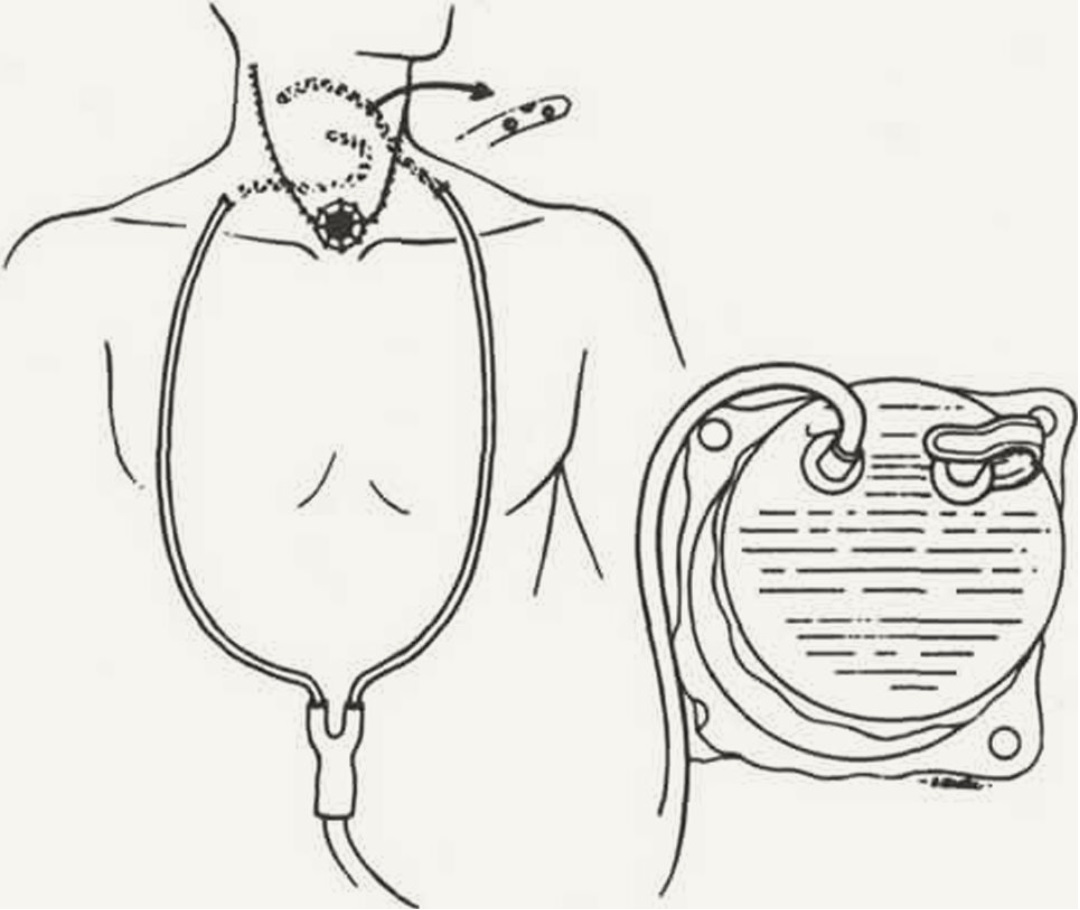
Closed drainage system for constant suction. Suction is maintained by a plastic container. The container serves as both suction source and receptacle for blooIt is emptied as required, and drainage tubes are left in the neck for approximately 3 days.
  1. Aphasia: impaired ability to understand or use commonly accepted words or symbols; interferes with ability to speak, write, or read; language center-usually left hemisphere (85% of population). Dysarthria is motor impairment-inability to articulate words.
    1. Types and pathophysiology:
      1. Receptive (sensory)-lesion usually Wernicke’s area of temporal lobe; difficulty understanding spoken word (auditory aphasia) or written word (visual aphasia).
      2. Expressive (motor)-difficulty expressing thoughts in speech or writing (motor aphasia); understands written and spoken words. Three types: anomic-fluent speech, but unable to name objects, qualities, and conditions; fluent-articulate and grammatically correct, but no content or meaning; nonfluent-unable to select, organize, and initiate speech (involves Brocas area of brain); may affect writing.
    2. Risk factors:
      1. Vascular disease of the brain (cerebrovascular accident).
      2. Alzheimer’s disease (degeneration).
      3. Tumor.
      4. Trauma.
    3. Analysis/nursing diagnosis:
      1. Impaired verbal communication related to cerebral cortex disorder.
      2. Powerlessness related to inability to express needs/concerns.
      3. Impaired social interaction related to difficulty communicating.
    4. Nursing care plan/implementation: Goal: assist with communication: patient does best when rested; small improvements will occur up to 1 year after injury (age is a factor). Strategies: Nonfluent -allow time to respond; support efforts to speak; acknowledge frustration of patient-anticipate needs when appropriate; use picture board or flashcards, pointing, to encourage communication; assess efforts to communicate with open-ended questions. Fluent-face the patient, speak slowly and distinctly, not loudly; use gestures, repeat instructions as needed; involve family in techniques to improve communication; acknowledge frustration.
    5. Evaluation/outcome criteria:
      1. Communication reestablished.
      2. Minimal frustration exhibited.
      3. Participates in speech therapy.

  2. Meniere’s disease: chronic, recurrent disorder of inner ear; attacks of vertigo, tinnitus, and vestibular dysfunction; lasts 30 minutes to full day; usually no pain or loss of consciousness.
    1. Pathophysiology: associated with excessive dilation of cochlear duct (unilateral) from overproduction or decreased absorption of endolymph (endolymphatic hydrops)-progressive sensorineural loss.
    2. Risk factors:
      1. Emotional or endocrine disturbance (diabetes mellitus).
      2. Spasms of internal auditory artery.
      3. Head trauma.
      4. Allergic reaction.
      5. High salt intake.
      6. Smoking.
      7. Ear infections.
    3. Assessment:
      1. Subjective data:
        1. Tinnitus (constant or intermittent).
        2. Headache; feeling of fullness or pressure in affected ear.
        3. True vertigo: sudden attacks; room appears to spin.
        4. Depression; irritability; withdrawal.
        5. Nausea with sudden head motion.
      2. Objective data:
        1. Impaired hearing, especially low tones.
        2. Change in gait; lack of coordination.
        3. Vomiting with sudden head motion.
        4. Nystagmus-during attacks.
        5. Diagnostic test: caloric (cold water in ear canal)-may precipitate attack; audiometry-loss of hearing.
    4. Analysis/nursing diagnosis:
      1. Risk for injury related to vertigo, lack of coordination.
      2. Auditory sensory/perceptual alteration related to progressive hearing loss.
      3. Anxiety related to uncertainty of treatment.
      4. Risk for activity intolerance related to sudden onset of vertigo.
      5. Sleep pattern disturbance related to tinnitus.
      6. Ineffective individual coping related to chronic disorder.
    5. Nursing care plan/implementation:
      1. Goal: provide safety and comfort during attacks.
        1. Activity: bed rest during attack; side rails up; lower to chair or floor if attack occurs while standing; assist with ambulation (sudden dizziness common).
        2. Position: recumbent; affected ear uppermost usually.
        3. Identify prodromal symptoms (aura, ear pressure, increased tinnitus).
      2. Goal: minimize occurrence of attacks.
        1. Give medications as ordered:
          1. Anticholinergics (oral or transdermal scopolamine, atropine, glycopyrrolate [Robinul]) to minimize GI symptoms.
          2. Antihistamines (dimenhydrinate [Dramamine], diphenhydramine HC1 [Benadryl]) to sedate vestibular system.
          3. Antiemetics and antivertigo agents (diazepam [Valium], meclizine HC1 [Antivert]).
          4. Diuretics may help (hydrochlorothiazide) to decrease endolymphatic fluid.
        2. Diet: low sodium (<2 g/day).
        3. Avoid precipitating stimuli: bright, glaring lights; noise; sudden jarring; turning head or eyes (stand in front of patient when talking).
      3. Goal: health teaching.
        1. No smoking (causes vasospasm) or alcoholic beverages (fluid retention, contraindicated with medications).
        2. Management of symptoms: play radio to mask tinnitus, particularly at night.
        3. Keep medication available at all times.
        4. Prepare for surgery if indicated (labyrinthectomy if hearing gone; or vestibular neurectomy to relieve vertigo and preserve hearing).
    6. Evaluation/outcome criteria:
      1. Decreased frequency of attacks.
      2. Complies with treatment regimen and restrictions (low-sodium diet, no smoking).
      3. Hearing preserved.
  1. Otosclerosis: disease of the bone of otic capsule; insidous, progressive deafness; most common cause of conductive deafness; cause unknown.
    1. Pathophysiology: formation of new spongy bone in labyrinth-fixation of stapes-prevention of sound transmission through ossicles to inner ear fluids.
    2. Risk factors:
      1. Heredity.
      2. Women, puberty to 45 years
      3. Pregnancy.
    3. Assessment:
      1. Subjective data:
        1. Difficulty hearing-gradual loss in both ears.
      2. Diagnostic tests:
        1. Rinne (tuning fork placed over mastoid bone)-reduced sound conduction by air and intensified by bone.
        2. Weber (tuning fork placed on top of head)-increased sound conduction to affected ear.
        3. Audiometry-diminished hearing ability.
    4. Analysis/nursing diagnosis:
      1. Auditory sensory/perceptual alteration related to hearing loss.
      2. Body image disturbance related to hearing aid.
      3. Ineffective individual coping related to grief reaction to loss.
      4. Impaired social interaction related to hearing loss.
    5. Nursing care plan/implementation, evaluation/ outcome criteria.

  2. Stapedectomy: removal of the stapes and replacing it with a prosthesis (steel wire, Teflon piston, or polyethylene); treatment for deafness due to otosclerosis, which fixes the stapes, preventing it from oscillating and transmitting vibrations to the fluids in the inner ear.
    1. Analysis/nursing diagnosis:
      1. Sensory/perceptual alteration related to edema and ear packing.
    2. Nursing care plan/implementation:
      1. Preoperative health teaching.
        1. Important to keep head in position ordered by physician postoperatively.
        2. Caution: sneezing, blowing nose (keep mouth open), vomiting, coughing-all of which increase pressure in eustachian tubes (blow one side gently).
        3. Breathing exercises.
      2. Postoperative:
        1. Goal: promote physical and psychological equilibrium.
          1. Position: as ordered by physician-varies according to preference.
          2. Activity: assist with ambulation; avoid rapid turning, which might increase vertigo.
          3. Dressings: check frequently; may change cotton pledget in outer ear.
          4. Give medications as ordered:
            1. Antiemetics.
            2. Analgesics.
            3. Antibiotics.
          5. Reassurance: reduction in hearing is normal; hearing may not immediately improve after surgery.
        2. Goal: health teaching.
          1. Ear care: keep covered outdoors; keep outer ear plug clean, dry, and changed.
          2. Avoid:
            1. Water in ear for 6 weeks:
              1. Use barrier when washing hair.
              2. Use two pieces of cotton; saturate outer piece with petroleum jelly.
            2. Pressure or vibration from loud noise, flying, or heavy lifting until advised by MD.
    3. Evaluation/outcome criteria:
      1. Hearing improves-evaluate 1 month postoperatively (may require hearing aid).
      2. Returns to work (usually 2 weeks after surgery).
      3. Continues medical supervision.
  1. Deafness: Hard of hearing-slight or moderate hearing loss that is serviceable for activities of daily living. Deaf-hearing is nonfunctional for activities of daily living.
    1. Risk factors:
      1. Conductive hearing losses (transmission deafness):
        1. Impacted cerumen (wax).
        2. Foreign body in external auditory canal.
        3. Defects (thickening, scarring) of eardrum.
        4. Otosclerosis of ossicles.
      2. Sensorineural hearing losses (perceptive or nerve deafness):
        1. Arteriosclerosis.
        2. Infectious diseases (mumps, measles, meningitis).
        3. Drug toxicities (quinine, streptomycin, neomycin SO4).
        4. Tumors.
        5. Head traumas.
        6. High-intensity noises.
      3. Central deafness:
        1. Tumors.
        2. Stroke (brain attack).
      4. Noise-induced or occupational noise hearing loss:
        1. Blast injury.
        2. Firearms.
        3. Loud music.
      5. Aging (presbycusis).
    2. Assessment-objective data:
      1. Inattentive or strained facial expression.
      2. Excessive loudness or softness of speech.
      3. Frequent need to clarify content of conversation or inappropriate responses.
      4. Tilting of head while listening.
      5. Lack of response when others speak.
    3. Analysis/nursing diagnosis:
      1. Auditory/sensory/perceptual alteration related to loss of hearing.
      2. Impaired social interaction related to deafness.
    4. Nursing care plan/implementation:
      1. Goal: maximize hearing ability and provide emotional support.
        1. Gain person’s attention before speaking; avoid startling.
        2. Provide adequate lighting so person can see you when you are speaking.
        3. Look at the person when speaking.
        4. Use nonverbal cues to enhance communication, e.g., writing, hand gestures, pointing.
        5. Speak slowly, distinctly; do not shout (excessive loudness distorts voice).
        6. If person does not understand, use different words; write it down.
        7. Use alternative communication system:
          1. Speech (lip) reading.
          2. Sign language.
          3. Hearing aid.
          4. Paper and pencil.
          5. Flash cards.
        8. Supportive, nonstressful environment; alert staff to patient’s hearing impairment.
      2. Goal: health teaching.
        1. Prepare for evaluative studies-audiogram.
        2. Appropriate community resources: National Association of Hearing and Speech Agencies for counseling services; National Association for the Deaf to assist with employment, education, legislation; Alexander Graham Bell Association for the Deaf, Inc., serves as information center for those working with the deaf; American Hearing Society provides educational information, employment services, social clubs.
        3. Use of hearing aid: care-(1) turn off aid when not using (open case at night to prevent draining battery); (2) keep extra battery available; (3) wash with mild soap and warm water; use pipe cleaner to remove wax; (4) do not wear aid in shower or with ear infection; (5) store same place every night and away from pets; (6) if aid does not work: check battery insertion, on-off switch, placement (if ear-molded aid), cleanliness.
        4. Safety precautions: when crossing street, driving.
    5. Evaluation/outcome criteria:
      1. Method of communication established.
      2. Achieves independence (use of Dogs for Deaf, special telephones, visual signals).
      3. Copes with lifestyle changes (minimal depression, anger, hostility).
  1. Glaucoma (acute and chronic): increased intraocular pressure; second most common cause of blindness.
    1. Pathophysiology:
      1. Acute (closed-angle)-impaired passage of aqueous humor into the circular canal of Schlemm due to closure of the angle between the cornea and the iris. Medical emergency; requires surgery.
      2. Chronic (open-angle)-degenerative changes in trabecular meshwork; local obstruction of aqueous humor between the anterior chamber and the canal. Most common; treated with medication (miotics, carbonic anhydrase inhibitors).
      3. Secondary-in some cases neovascularization (new vessels) may form; blocks passage of aqueous humor (uveitis, trauma, drugs, diabetes, retinal vein occlusion).
      4. Untreated: imbalance between rate of secretion of intraocular fluids and rate of absorption of aqueous humor-increased intraocular pressures-decreased peripheral vision-corneal edema-halos and blurring of vision-blindness.
    2. Risk factors-unknown, but associated with:
      1. Emotional disturbances.
      2. Hereditary factors.
      3. Allergies.
      4. Vasomotor disturbances.
    3. Assessment:
      1. Subjective data:
        1. Acute (closed-angle):
          1. Pain: severe, in and around eyes.
          2. Rainbow halos around lights.
          3. Blurring of vision.
          4. Nausea, vomiting.
        2. Chronic (open-angle):
          1. Eyes tire easily.
          2. Loss of peripheral vision.
          3. Dull, morning headache.
      2. Objective data:
        1. Corneal edema.
        2. Decreased peripheral vision.
        3. Increased cupping of optic disc.
        4. Tonometry-pressures >22 mm Hg.
        5. Pupils: dilated.
        6. Redness of eye.
    4. Analysis/nursing diagnosis:
      1. Visual sensory/perceptual alterations related to increased intraocular pressure.
      2. Pain related to sudden increase in intraocular pressure.
      3. Risk for injury related to blindness.
      4. Impaired physical mobility related to impaired vision.
    5. Nursing care plan/implementation:
      1. Goal: reduce intraocular pressure.
        1. Activity: bed rest.
        2. Position: semi-Fowler’s.
        3. Medications as ordered:
          1. Miotics (pilocarpine, carbachol); may not be effective with intraocular pressure (IOP) >40 mm Hg.
          2. Carbonic anhydrase inhibitors (acetazolamide [Diamox]).
          3. Anticholinesterase (demecarium bromide [Humorsol]) to facilitate outflow of aqueous humor.
          4. Ophthalmic beta blockers (timolol) to decrease IOP.
      2. Goal: provide emotional support.
        1. Place personal objects within field of vision.
        2. Assist with activities.
        3. Encourage verbalization of concerns, fears of blindness, loss of independence.
      3. Goal: health teaching.
        1. Prevent increased IOP by avoiding:
          1. Anger, excitement, worry.
          2. Constrictive clothing.
          3. Heavy lifting.
          4. Excessive fluid intake.
          5. Atropine or other mydriatics that cause dilation.
          6. Straining at stool.
          7. Eye strain.
        2. Relaxation techniques; stress management if indicated.
        3. Prepare for surgical intervention, if ordered: laser trabeculoplasty, trabeculectomy (filtering), laser peripheral iridotomy.
        4. Medications: purpose, dosage, frequency; eyedrop installation-(1) wash hands; (2) head back, expose conjunctiva of lower lid, instill in center without touching eyelashes or eye; (3) close eyes gently, apply slight pressure to corner of eye to decrease systemic absorption; (4) wait at least 2 minutes before instilling a second eyedrop medication; have extra bottle in case of breakage or loss.
        5. Activity: moderate exercise-walking.
        6. Safety measures: eye protection (shield or glasses); Medic Alert band or tag; avoid driving 1-2 hours after instilling miotics.
        7. Community resources as necessary.
    6. Evaluation/outcome criteria:
      1. Eyesight preserved if possible
      2. Intraocular pressure lowered (<22 mm Hg).
      3. Continues medical supervision for life-reports reappearance of symptoms immediately.
  1. Cataract: developmental or degenerative opacification of the crystalline lens.
    1. Risk factors:
      1. Aging (most common).
      2. Trauma (x-rays, infrared or possibly ultraviolet exposure).
      3. Systemic disease (diabetes).
      4. Congenital defect.
      5. Drug effects (corticosteroids).
    2. Assessment:
      1. Subjective data-vision: blurring, loss of acuity (see best in low-lit conditions); distortion; diplopia; photophobia.
      2. Objective data:
        1. Blindness: unilateral or bilateral (particularly in congenital cataracts).
        2. Loss of red reflex; gray or cloudy white opacity of lens.
    3. Analysis/nursing diagnosis:
      1. Visual sensory/perceptual alterations related to opacity of lens.
      2. Risk for injury related to accidents.
      3. Social isolation related to impaired vision.

  2. Cataract removal: removal of opacified lens because of loss of vision; extracapsular cataract extraction followed by intraocular lens (IOL) insertion is procedure of choice.
    1. Nursing care plan/implementation:
      1. Preoperative:
        1. Goal: prepare for surgery (ambulatory center). Antibiotic drops or ointment, mydriatic eyedrops as ordered; note dilation of pupils; avoid glaring lights; usually done under local anesthetic with sedation.
        2. Goal: health teaching. Postoperative expectations: do not rub, touch, or squeeze eyes shut after surgery; eye patches will be on; assistance will be given for needs; overnight hospitalization not required unless complications occur; mild iritis usually occurs.
      2. Postoperative:
        1. Goal: reduce stress on the sutures and prevent hemorrhage.
          1. Activity: ambulate as ordered, usually soon after surgery; generally discharged few hours after surgery.
          2. Position: flat or low Fowler's; on hack or turn to nonoperative side 3-4 weeks, because turning to operative side increases pressure.
          3. Avoid activities that increase IOP: straining at stool, vomiting, coughing, brushing teeth, brushing hair, shaving, lifting objects over 20 lb, bending, or stooping; wear glasses or shaded lens during day, eyeshield at night.
          4. Provide mouthwash, hair care, personal items within easy reach, “stepin” slippers to avoid bending over.
        2. Goal: promote psychological well-being with elderly, frequent contacts to prevent sensory deprivation.
        3. Goal: health teaching.
          1. If intraocular lens not inserted, prescriptive glasses may be used (cataract glasses); explain about magnification, perceptual distortion, blind areas in peripheral vision; guide through activities with glasses; need to look through central portion of lens and turn head to side when looking to the side to decrease distortion.
          2. Eye care: instillation of eyedrops (mydriatics and carbonic anhydrase inhibitors) to prevent glaucoma and adhesions if IOL not inserted; with IOL, steroid-antibiotic use
          3. Signs/symptoms of infection (redness, pain, edema, drainage); iris prolapse (bulging or pear-shaped pupil); hemorrhage (sharp eye pain, halfmoon of blood).
          4. Avoid: heavy lifting; potential eye trauma.
    2. Evaluation/outcome criteria:
      1. Vision restored.
      2. No complications (severe eye pain, hemorrhage).
      3. Performs self-care activities (instills eyedrops).
      4. Returns for follow-up ophthalmology care-recognizes symptoms requiring immediate attention.
  1. Retinal detachment: separation of neural retina from underlying retinal pigment epithelium.
    1. Risk factors:
      1. Trauma.
      2. Degeneration.
    2. Assessment:
      1. Subjective data:
        1. Flashes of light before eyes.
        2. Vision: blurred, sooty (sudden onset); sensation of floating particles; blank areas of vision.
      2. Objective data-ophthalmic exam: retina is grayish in area of tear; bright red, horseshoe shaped tear; B-scan ultrasonography.
    3. Analysis/nursing diagnosis:
      1. Visual sensory/perceptual alteration related to blurred vision.
      2. Anxiety related to potential loss of vision.
      3. Risk for injury related to blindness.
    4. Nursing care plan/implementation:
      1. Preoperative:
        1. Goal: reduce anxiety and prevent further detachment.
          1. Encourage verbalization of feelings; answer all questions; reinforce physician’s explanation of surgical procedures.
          2. Activity: bed rest; eyes usually covered to promote rest and maintain normal position of retina; side rails up.
          3. Position: according to location of retinal tear; involved area of eye should be in a dependent position.
          4. Give medications as ordered: cycloplegic or mydriatics to dilate pupils widely and decrease intraocular movement.
          5. Relaxing diversion: conversation, music.
        2. Goal: health teaching. Prepare for surgical intervention (often combination used):
          1. Cryopexy or cryotherapy-supercooled probe is applied to the sclera, causing a scar, which pulls the choroid and retina together.
          2. Laser photocoagulation-a beam of intense light from a carbon arc is directed through the dilated pupil onto the retina; seals hole if retina not detached.
          3. Scleral buckling-the sclera is resected or shortened to enhance the contact between the choroid and retina; frequently combined with cryopexy.
          4. Banding or encirclement-silicone band or strap is placed under the extraocular muscles around the globe.
          5. Pneumatic retinopexy-instillation of expandable gas or oil to tamponade tear.
      2. Postoperative:
        1. Goal: reduce intraocular stress and prevent hemorrhage.
          1. Position: flat or low Fowler’s; sandbags may be used to position head; turn to nonoperative side if allowed, retinal tear dependent; special positions may be prone, side-lying, or sitting with face down on table if gas or oil bubble injected; position may be restricted 4-8 days.
          2. Activity: bed rest; decrease intraocular pressure by not stooping or bending and avoiding prone position.
          3. Give medications as ordered:
            1. Cycloplegics (atropine).
            2. Antibiotics.
            3. Corticosteroids to reduce eye movements and inflammation and prevent infection.
          4. ROM-isometric, passive; elastic stockings to avoid thrombus related to immobility.
        2. Goal: support coping mechanisms.
          1. Plan all care with patient.
          2. Encourage verbalization of feelings, fears.
          3. Encourage family interaction.
          4. Diversional activities.
        3. Goal: health teaching.
          1. Eye care: eye patch or shield at night for about 2 weeks to prevent touching eye while asleep; dark glasses; avoid rubbing, squeezing eyes.
          2. Limitations: no reading, needlework for 3 weeks, no physical exertion for 6 weeks; OK to watch TV, walk, except with bubble restrictions.
          3. Medications: dosage, frequency, purpose, side effects: avoid nonprescription medications.
          4. Signs of redetachment: flashes of light, increase in “floaters,” blurred vision, acute eye pain.
    5. Evaluation/outcome criteria:
      1. Vision restored.
      2. No further detachment-recognizes signs and symptoms.
      3. No injury occurs-accepts limitations.

  2. Blindness: legally defined as vision less than 20/200 with the use of corrective lenses, or a visual field of no greater than 20 degrees; greatest incidence after 65 years.
    1. Risk factors:
      1. Glaucoma.
      2. Cataracts.
      3. Macular degeneration.
      4. Diabetic retinopathy.
      5. Atherosclerosis.
      6. Trauma.
    2. Analysis/nursing diagnosis:
      1. Visual sensory/perceptual alteration related to blindness.
      2. Impaired social interaction related to loss of sight.
      3. Risk for injury related to visual impairment.
      4. Self-care deficit related to visual loss.
    3. Nursing care plan/implementation:
      1. Goal: promote independence and provide emotional support.
        1. Familiarize with surroundings; encourage use of touch.
        2. Establish communication lines; answer questions.
        3. Deal with feelings of loss, overprotectiveness by family members.
        4. Provide diversional activities: radio, CDs, talking books, tapes.
        5. Encourage self-care activities; allow voicing of frustrations when activity is not done to satisfaction (spilling or misplacing something), to decrease anger and discouragement.
      2. Goal: facilitate activities of daily living.
        1. Eating:
          1. Establish routine placement for tableware, plate, glass.
          2. Help person mentally visualize the plate as a clock or compass (“3 o’clock” or “east”).
          3. Take person’s hand and guide the fingertips to establish spatial relationship.
        2. Walking:
          1. Have person hold your forearm: walk a half step in front.
          2. Tell the person when approaching stairs, curb, incline.
        3. Talking:
          1. Speak when approaching person; tell them before you touch them.
          2. Tell them who you are and what you will be doing.
          3. Do not avoid words such as “see” or discussing the appearance of things.
      3. Goal: health teaching.
        1. Accident prevention in the home.
        2. Community resources:
          1. Voluntary agencies:
            1. American Foundation for the Blind-provides catalogs of devices for visually handicapped.
            2. National Society for the Prevention of Blindness-comprehensive educational programs and research.
            3. Recording for the Blind, Inc.-provides recorded educational books on free loan.
            4. Lion’s Club.
            5. Catholic charities.
            6. Salvation Army.
          2. Government agencies:
            1. Social and Rehabilitation Service-counseling and placement services.
            2. Veterans Administration-screening and pensions.
            3. State Welfare Department, Division for the Blind-vocational.
    4. Evaluation/outcome criteria:
      1. Acceptance of disability-participates in selfcare activities, remains socially involved.
      2. Regains independence with rehabilitation.
  1. Traumatic injuries to the brain
    1. Primary trauma:
      1. Concussion- transient disorder due to injury in which there is brief loss of consciousness due to paralysis of neuronal function; recovery is usually total.
      2. Contusion- structural alteration of brain tissue characterized by extravasation of blood cells (bruising); injury may occur on side of impact or on opposite side (when cranial contents shift forcibly within the skull with impact).
      3. Laceration- tearing of brain tissue or blood vessels due to a sharp bone fragment or object or tearing force.
      4. Fracture- linear (may result in epidural bleed); comminuted or depressed (may tear dura and result in CSF leak); basilar (most serious). Basilar skull fracture may result in meningitis or brain abscess; bleeding from nose or ears; CSF present in drainage; bruising over mastoid process (Battles sign) and periorbital ecchymosis (raccoon eyes).
    2. Secondary trauma (response to primary trauma):
      1. Hematomas:
        1. Subdural- blood from ruptured or torn vein collects between arachnoid and dura; may be acute, subacute, or chronic.
        2. Extradural (epidural)-blood clot located between dura mater and inner surface of skull; most often from tearing of middle meningeal artery; emergency condition.
      2. Increased intracranial pressure
    3. Mechanisms of injury:
      1. Deformation (blow to the head).
      2. Acceleration-deceleration (coup-contrecoup)-forward and rebounding motion.
      3. Rotation (tension, stretching, shearing force).
    4. Pathophysiology of impaired CNS functioning:
      1. Depressed neuronal activity in reticular activating system-depressed consciousness
      2. Depressed neuronal functioning in lower brain stem and spinal cord-depression of reflex activity-decreased eye movements, unequal pupils-decreased response to light stimuli-widely dilated and fixed pupils.
      3. Depression of respiratory center-altered respiratory pattern-decreased rate respiratory arrest.
    5. Risk factors: accidents-automobile, industrial and home, motorcycle, military.
    6. Assessment:
      1. Objective data:
        1. Headache.
        2. Dizziness, loss of balance.
        3. Double vision.
        4. Nausea.
      2. Objective data:
        1. Laceration or abrasion around face or head; profuse bleeding from scalp (highly vascular, poor vasoconstrictive abilities).
        2. Drainage from ears or nose (serosanguineous).
        3. Projectile vomiting, hematemesis.
        4. Vital signs indicating increased intracranial pressure
        5. Neurological exam:
          1. Altered level of consciousness; a numerical assessment, such as the Glasgow Coma Scale, may be used. The lower the score, the poorer the prognosis, generally.
          2. Pupils-equal, round, react to light; or unequal, dilated, unresponsive to light.
          3. Extremities-paresis or paralysis.
          4. Reflexes-hypotonia or hypertonia; Babinski present (flaring of great toe when sole is stroked).
    7. Analysis/nursing diagnosis:
      1. Altered thought processes related to brain trauma.
      2. Sensory/perceptual alteration related to depressed neuronal activity.
      3. Risk for injury related to impaired CNS functioning.
      4. Risk for aspiration related to respiratory depression.
      5. Self-care deficit related to altered level of consciousness.
      6. Ineffective breathing pattern related to CNS trauma.
    8. Nursing care plan/implementation:
      1. Goal: sustain vital functions and minimize or prevent complications.
        1. Patent airway: endotracheal tube or tracheostomy may be ordered.
        2. Oxygen: as ordered (hypoxia increases cerebral edema).
        3. Be Position: semiprone or prone (coma position) with head level to prevent aspiration (keep off back); turn side to side to prevent stasis in lungs.
        4. Vital signs as ordered.
        5. Neurological check: pupils, level of consciousness, muscle strength; report changes.
        6. Seizure precautions: padded side rails.
        7. Medications as ordered:
          1. Steroids (dexamethasone [Decadron]).
          2. Anticonvulsants (phenytoin [Dilantin], phenobarbital).
          3. Analgesics (morphine contraindicated).
        8. Cooling measures or hypothermia to reduce elevated temperature.
        9. Assist with diagnostic tests:
          1. Lumbar puncture (contraindicated with increased intracranial pressure).
          2. Electroencephalogram (EEG).
        10. Diet: NPO for 24 hours, progressing to clear liquids if awake.
        11. Fluids: IVs; nasogastric tube feedings; I&O.
        12. Monitor blood chemistries: sodium imbalance common with head injuries.
      2. Goal: provide emotional support and use comfort measures.
        1. Comfort: skin care, oral hygiene; sheepskins; wrinkle-free linen.
        2. Eyes: lubricate q4h with artificial tears if periocular edema present.
        3. ROM-passive, active; physical therapy as tolerated.
        4. Avoid restraints.
        5. Encourage verbalization of concerns about changes in body image, limitations.
        6. Encourage family communication.
    9. Evaluation/outcome criteria:
      1. Alert, oriented-no residual effects (cognitive processes intact).
      2. No signs of increased intracranial pressure (decreased respirations, increased systolic pressure with widening pulse pressure, bradycardia).
      3. No paralysisregains motor/sensory function.
      4. Resumes self-care activities.

Levels of Consciousness

Stage Characteristics

Alertness


Aware of time and place


Automatism


Aware of time and place but demonstrates abnormality of mood (euphoria to irritability)


Confusion


Inability to think and speak in coherent manner; responds to verbal requests but is unaware of time and place


Delirium


Restlessness and violent activity; may not comply with verbal instructions


Stupor


Quiet and uncommunicative; may appear conscious-sits or lies with glazed look; unable to respond to verbal instructions; bladder and rectal incontinence may occur


Semicoma


Unresponsive to verbal instructions but responds to vigorous or painful stimuli


Coma


Unresponsive to vigorous or painful stimuli


Glasgow Coma Scale

Best eye-opening response

Purposeful and spontaneous:

  • To voice
  • To pain

No response

Untestable

Best verbal response

  • Oriented
  • Disoriented
  • Inappropriate words
  • Incomprehensible sounds
  • Untestable

Best motor response

  • Obeys commands
  • Localizes pain
  • Withdraws to pain
  • Extension to pain
  • No response
  • Untestable
  1. Increased intracranial pressure (ICP): intracranial hypertension associated with altered states of consciousness.
    1. Pathophysiology: increases in intracranial blood volume, cerebrospinal fluid, or brain tissue mass-increased intracranial pressure-impaired neural impulse transmission cellular anoxia, atrophy.
    2. Risk factors:
      1. Congenital anomalies (hydrocephalus).
      2. Space-occupying lesions (abscesses or tumors).
      3. Trauma (hematomas or skull fractures).
      4. Circulatory problems (aneurysms, emboli).
      5. Inflammation (meningitis, encephalitis).
    3. Assessment:
      1. Subjective data:
        1. Headache (early, but nonspecific symptom).
        2. Nausea.
        3. Visual disturbance (diplopia).
      2. Objective data:
        1. Changes in level of consciousness (early sign).
        2. Pupillary changes-unequal, dilated, and unresponsive to light (late sign).
        3. Vital signs-changes are variable.
          1. Blood pressure-gradual or rapid elevation, widened pulse pressure.
          2. Pulse-bradycardia, tachycardia; significant sign is slowing of pulse as blood pressure rises.
          3. Respirations-pattern changes(Cheyne-Stokes, apneusis, Biot’s), deep and sonorous; hiccups.
          4. Temperature-moderate elevation.
        4. Projectile vomiting (more common in children).
        5. Diagnostic test: Head CT-structural changes.
    4. Analysis/nursing diagnosis:
      1. Altered cerebral tissue perfusion related to increased intracranial pressure.
      2. Altered thought processes related to cerebral anoxia.
      3. Ineffective breathing pattern related to compression of respiratory center.
      4. Risk for aspiration related to unconsciousness.
      5. Self-care deficit related to altered level of consciousness.
      6. Impaired physical mobility related to abnormal motor responses.
    5. Nursing care plan/implementation:promote adequate oxygenation and limit further impairment.
      1. Vital signs: report changes immediately.
      2. Patent airway; keep alkalotic, to prevent increased intracranial pressure from elevated CO2; hyperventilate if necessary.
      3. Give medications as ordered:
        1. Hyperosmolar diuretics (mannitol, urea) to reduce brain swelling.
        2. Steroids (dexamethasone [Decadron]) for anti-inflammatory action.
        3. Antacids or H2 antagonist to prevent stress ulcer.
      4. B Position: head of bed elevated 30 degrees.
      5. Fluids: restrict; strict I&O.
      6. Cooling measures to reduce temperature, because fever increases ICP.
      7. Prepare for surgical intervention.
    6. Evaluation/outcome criteria:
      1. No irreversible brain damage-regains consciousness.
      2. Resumes self-care activities.

  2. Craniotomy: excision of a part of the skull (burr hole to several centimeters) for exploratory purpose and biopsy; to remove neoplasms, evacuate hematomas or excess fluid, control hemorrhage, repair skull fractures, remove scar tissue, repair or excise aneurysms, and drain abscesses; produces minimal neurological deficit.
    1. Analysis/nursing diagnosis:
      1. Altered cerebral tissue perfusion related to edema.
      2. Altered thought processes related to disorientation.
      3. Self-care deficit related to continued neurological impairment.
    2. Nursing care plan/implementation:
      1. Preoperative:
        1. Goal: obtain baseline measures.
          1. Vital signs.
          2. Level of consciousness.
          3. Mental, emotional status.
          4. Pupillary reactions.
          5. Motor strength and functioning.
        2. Goal: provide psychological support: listen; give accurate, brief explanations.
        3. Goal: prepare for surgery.
          1. Cut hair; shave scalp (usually done in surgery); save hair if patient/family desire.
          2. Cover scalp with clean towel.
          3. Insert indwelling Foley catheter as ordered.
      2. Postoperative:
        1. Goal: prevent complications and limit further impairment.
          1. Vital signs (indications of complications):
            1. Decreased blood pressure-shock.
            2. Widened pulse pressure-increased ICP.
            3. Respiratory failure-compression of medullary respiratory centers.
            4. Hyperthermia-disturbance of heat-regulating mechanism; infection.
          2. Neurological:
            1. Pupils-ipsilateral dilation (increased ICP), visual disturbances.
            2. Altered level of consciousness.
            3. Altered cognitive or emotional status-disorientation common.
            4. Motor function and strength- hypertonia, hypotonia, seizures.
          3. Blood gases, to monitor adequacy of ventilation.
          4. Dressings: check frequently; aseptic technique; reinforce as necessary.
          5. Observe for:
            1. CSF leakage (glucose-positive drainage from nose, mouth, ears)-report immediately.
            2. Periorbital edema-apply light ice compresses as necessary remove crusts from eyelids.
          6. Check integrity of seventh cranial nerve (facial)-incomplete closure of eyelids.
          7. Position:
            1. Supratentorial surgery (cerebrum)-semi-Fowler’s (30-degree elevation); may not lie on operative side.
            2. Infratentorial (brain stem, cerebellum)-flat in bed (prone); may turn to either side but not onto back.
          8. Fluids: NPO until bowel sounds return, alert, and intact gag, swallow, and cough reflexes present.
          9. Medications as ordered:
            1. Osmotic diuretics (mannitol).
            2. Corticosteroids (dexamethasone [Decadron]).
            3. Mild analgesics (do not mask neurological or respiratory depression).
            4. Stool softeners to prevent constipation and straining.
          10. Orient frequently to person, time, place-to reduce restlessness, confusion.
          11. Side rails up for safety.
          12. Avoid restraints (may increase agitation and ICP).
          13. Ice bags to head to reduce headache.
          14. Activity: assist with ambulation.
        2. Goal: provide optimal supportive care.
          1. Cover scalp once dressings are removed (scarves, wigs).
          2. Deal realistically with neurological deficits-facilitate acceptance, adjustment, independence.
        3. Goal: health teaching.
          1. Prepare for physical, occupational, or speech therapy, as needed.
          2. Activities of daily living.
    3. Evaluation/outcome criteria:
      1. Regains consciousness-is alert, oriented.
      2. Resumes self-care activities within limits of neurological deficits.
  1. Epilepsy: seizure disorder characterized by sudden transient aberration of brain function; associated with motor, sensory, autonomic, or psychic disturbances.
    1. Seizure: involuntary muscular contraction and disturbances of consciousness from abnormal electrical activity.
      1. Brain injury.
      2. Infection (meningitis, encephalitis).
      3. Water and electrolyte disturbances.
      4. Hypoglycemia.
      5. Tumors.
      6. Vascular disorders (hypoxia or hypocapnia).
    2. Risk factors:
    3. Generalized seizures:
      1. Tonic-clonic (grand mal) seizures:
        1. Pathophysiology: increased excitability of a neuron-possible activation of adjacent neurons-synchronous discharge of impulses-vigorous involuntary sustained muscle spasms (tonic contractions). Onset of neuronal fatigue- intermittent muscle spasms (clonic contractions)-cessation of muscle spasms-fatigue.
        2. Assessment:
          1. Subjective data- aura: flash of light; peculiar smell, sound; feelings of fear; euphoria.
          2. Objective data:
            1. Convulsive stage- tonic and clonic muscle spasms, loss of consciousness, breath holding, frothing at mouth, biting of tongue, urinary or fecal incontinence; lasts 2-5 minutes.
            2. Postconvulsion- headache, fatigue (postictal sleep), malaise, vomiting, sore muscles, choking on secretions, aspiration.
      2. Absence (petit mal) seizures:
        1. Pathophysiology: unknown etiology, momentary loss of consciousness (10-20 seconds); usually no recollection of seizure; resumes previously performed action.
        2. Assessment- objective data:
          1. Fixation of gaze; blank facial expression.
          2. Flickering of eyelids.
          3. Jerking of facial muscle or arm.
      3. Minor motor seizures:
        1. Myoclonic -involuntary “lightning-like” jerking contraction of major muscles; may throw person to the floor; no loss of consciousness.
        2. Atonic -brief, total loss of muscle tone; person falls to the floor; loss of consciousness (common in children).
    4. Partial (focal) seizures:
      1. Partial motor: arises from region in motor cortex (posterior frontal lobe); most commonly begins in upper extremities, spreading to face and lower extremity (jacksonian march); noting progression is important in identifying area of cortex involved.
      2. Partial sensory: sensory symptoms occur with partial seizure activity; varies with region in brain; transient.
      3. Partial complex (psychomotor): arises out of anterior temporal lobe; frequently begins with an aura; characteristic feature is automatism (lip smacking, chewing, patting body, picking at clothes); lasts from 2-3 minutes to 15 minutes; do not restrain.
    5. Analysis/nursing diagnosis:
      1. Risk for injury related to convulsive disorder.
      2. Anxiety related to sudden loss of consciousness.
      3. Self-esteem disturbance related to chronic illness.
      4. Impaired social interaction related to selfconsciousness.
    6. Nursing care plan/implementation (generalized seizures):
      1. Goal: prevent injury during seizure.
        1. Do not force jaws open during convulsion.
        2. Do not restrict limbs-protect from injury; place something soft under head (towel, jacket, hands).
        3. Loosen constrictive clothing.
        4. Note time, level of consciousness, type and duration of seizure.
      2. Goal: postseizure care:
        1. Turn on side to drain saliva and facilitate breathing.
        2. Suction as necessary.
        3. Orient to time and place.
        4. Oral hygiene if tongue or cheek injured.
        5. Check vital signs, pupils, level of consciousness.
      3. Goal: prevent or reduce recurrences of seizure activity.
        1. Encourage patient to identify precipitating factors.
        2. Moderation in diet and exercise.
        3. Medications as ordered: phenytoin (Dilantin); phenobarbital; carbamazepine (Tegretol); primidone (Mysoline); valproate (Depacon).
      4. Goal: health teaching.
        1. Medications:
          1. Actions, side effects (apathy, ataxia, hyperplasia of gums).
          2. Complications with sudden withdrawal (status epilepticus -continuous seizure activity; give diazepam per order, O 2).
        2. Attitude toward life and treatment; adhere to medication program.
        3. Clarify misconceptions, fears-especially about insanity, bad genes.
        4. Maintain activities, interests-expect no driving until seizure free for period of time specified by state Department of Motor Vehicles.
        5. Avoid: stress; lack of sleep; emotional upset; alcohol.
        6. Relaxation techniques; stress-management techniques.
        7. Use Medic Alert band or tag.
        8. Appropriate community resources.
    7. Evaluation/outcome criteria:
      1. Avoids precipitating stimuli-achieves seizure control.
      2. Complies with medication regimen.
      3. Retains independence.

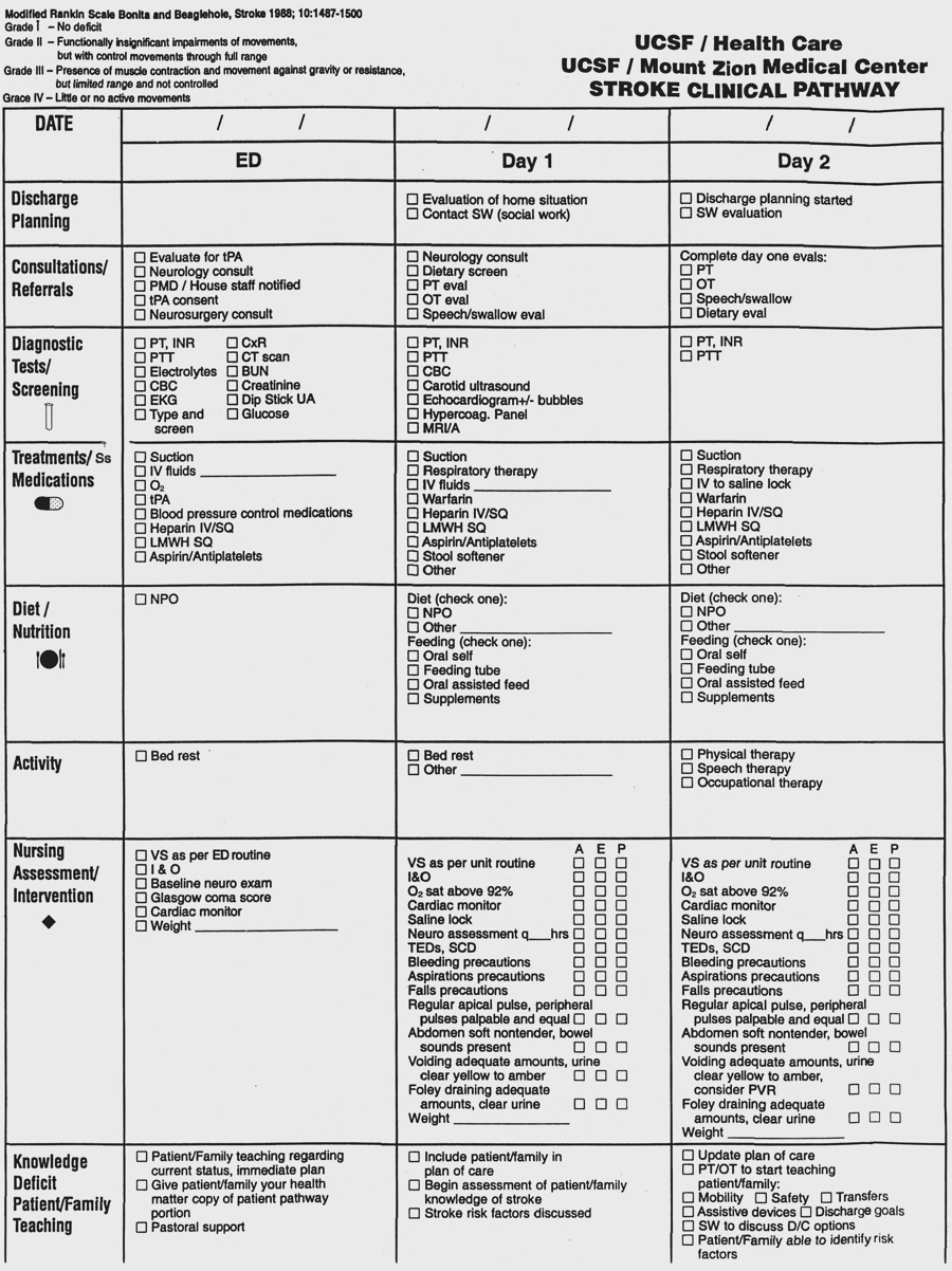
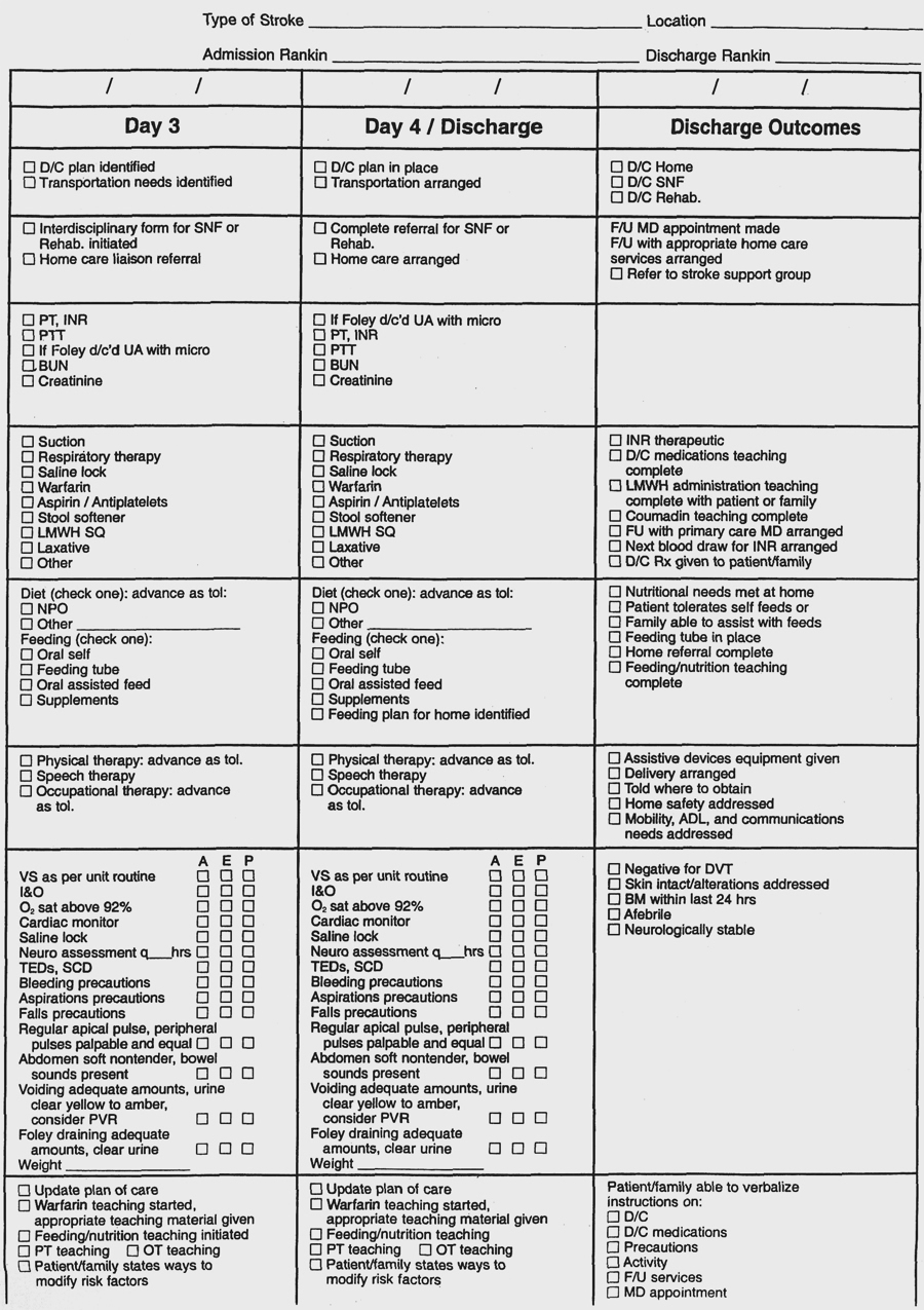
  2. Transient Ischemic Attacks (TIAs): temporary, complete, or relatively complete cessation of cerebral blood flow to a localized area of brain, producing symptoms (2-30 minutes) ranging from weakness (“drop attacks”) and numbness to monocular blindness; an important precursor to strokSurgical intervention includes carotid endarterectomy; most common postoperative cranial nerve damage causes vocal cord paralysis or difficulty managing saliva and tongue deviation (cranial nerves VII, X, XI, XII); usually temporary; stroke may also occur.
  1. Ischemic stroke (CVA, brain attack): brain lesion resulting from damage to blood vessels supplying brain (middle cerebral artery most common).
    1. Pathophysiology: reduced or interrupted blood flow-interruption of nerve impulses down corticospinal tract-decreased or absent voluntary movement on one side of the body (fine movements are more affected than coarse movements); later, autonomous reflex activity-spasticity and rigidity of muscles.
    2. Risk factors:
      1. Cerebral thrombosis (most common), embolism, hemorrhage.
      2. Prior ischemic episodes (TIAs).
      3. Hypertension.
      4. Oral contraceptives.
      5. Emotional stress.
      6. Family history.
      7. Age.
      8. Diabetes mellitus.
    3. Assessment:
      1. Subjective data:
        1. Weakness: sudden or gradual loss of movement of extremities on one side.
        2. Difficulty forming words.
        3. Difficulty swallowing (dysphagia).
        4. Nausea, vomiting.
        5. History of TIAs.
      2. Objective data:
        1. Vital signs:
          1. BP-elevated; widened pulse pressure.
          2. Temperature-elevated.
          3. Pulse-normal, slow.
          4. Respirations-tachypnea, altered pattern; deep; sonorous.
        2. Neurological:
          1. Altered level of consciousness.
          2. Pupils-unequal; vision-homonymous hemianopia.
          3. Ptosis of eyelid, drooping mouth.
          4. Paresis or paralysis (hemiplegia).
          5. Loss of sensation and reflexes.
          6. Incontinence of urine or feces.
          7. Aphasia
    4. Analysis/nursing diagnosis:
      1. Impaired physical mobility related to hemiplegia.
      2. Impaired swallowing related to paralysis.
      3. Impaired verbal communication related to aphasia.
      4. Risk for aspiration related to unconsciousness.
      5. Sensory/perceptual alterations related to altered cerebral blood flow, visual field blindness.
      6. Altered thought processes related to cerebral edema.
      7. Self-care deficit related to paresis or paralysis.
      8. Body image disturbance related to hemiplegia.
      9. Total incontinence related to interruption of normal nerve transmission.
      10. Impaired social interaction related to aphasia or neurological deficit.
      11. Risk for impaired skin integrity related to immobility.
      12. Unilateral neglect related to cerebral damage
    5. Nursing care plan/implementation:
      1. Goal: reduce cerebral anoxia.
        1. Patent airway:
          1. Oxygen therapy as ordered; suctioning to prevent aspiration.
          2. Turn, cough, deep breathe q2h due to high incidence of aspiration pneumonia.
        2. Activity: bed rest, progressing to out of bed as tolerated.
        3. Position:
          1. Maximize ventilation.
          2. Support with pillows when on side; use hand rolls and arm slings as ordered.
      2. Goal: promote cardiovascular function and maintain cerebral perfusion.
        1. Vital signs; neurological checks.
        2. Medications as ordered:
          1. Antiplatelet aggregation inhibitors (ASA, ticlopidine, dipyridamole [Persantine]).
          2. Antihypertensives to prevent rupture.
          3. Anticoagulants to prevent thrombus.
        3. Fluids: IVs to prevent hemoconcentration; I&O; weigh daily.
        4. ROM exercises to prevent contractures, muscle atrophy, phlebitis.
        5. Skin care and position changes to prevent decubiti.
      3. Goal: provide for emotional relaxation.
        1. Identify grief reaction to changes in body image.
        2. Encourage expression of feelings, concerns.
      4. Goal: patient safety.
        1. Identify existence of homonymous hemianopia (visual field blindness) and agnosia (disturbance in sensory information).
        2. Use side rails and assist as needed.
        3. Remind to walk slowly, take adequate rest periods, ensure good lighting, look where patient is going.
      5. Goal: health teaching.
        1. Exercise routines.
        2. Diet: self-feeding, but assist as needed.
        3. Resumption of self-care activities.
        4. Use of supportive devices; transfer techniques.
    6. Evaluation/outcome criteria:
      1. No complications (pneumonia).
      2. Regains functional independence-resumes self-care activities.
      3. Return of control over body functions (bowel, bladder, speech).


  1. Bacterial meningitis.

  2. Encephalitis (also includes aseptic meningitis): inflammation of the brain and its coverings due to direct viral invasion, which usually results in a lengthy coma.
    1. Pathophysiology: brain tissue injury- release of enzymes that increase vascular dilation, capillary permeability-edema formation-increased intracranial pressure-depression of CNS function.
    2. Risk factors:
      1. Arboviruses.
      2. Enteroviruses (poliovirus, echovirus).
      3. Herpesvirus.
      4. Varicella-zoster (chickenpox).
      5. Postinfection complication (measles, mumps, smallpox).
    3. Assessment:
      1. Subjective data:
        1. Headache-severe.
        2. Fever-sudden.
        3. Nausea, vomiting.
        4. Sensitivity to light (photophobia).
        5. Difficulty concentrating.
      2. Objective data:
        1. Altered level of consciousness.
        2. Nuchal rigidity.
        3. Tremors; facial weakness.
        4. Nystagmus.
        5. Elevated temperature.
        6. Diagnostic test: lumbar puncture-fluid cloudy; increased neutrophils, protein.
        7. Lab data: blood-slight to moderate leukocytosis (about 14,000).
    4. Analysis/nursing diagnosis:
      1. Self-care deficit related to altered level of consciousness.
      2. Risk for injury related to coma.
      3. Sensory/perceptual alteration related to brain tissue injury.
      4. Altered thought processes related to increased intracranial pressure.
    5. Nursing care plan/implementation:
      1. Goal: support physical and emotional relaxation.
        1. Vital signs; neurological signs as ordered.
        2. Seizure precautions.
        3. Position: to maintain patent airway; prevent contractures; ROM.
        4. Medications as ordered:
          1. Analgesics for headache and neck pain.
          2. Antipyretics for fever.
          3. Antivirals.
          4. Anticonvulsants for seizures.
          5. Antibiotics for infection in aseptic meningitis.
          6. Osmotic diuretics to reduce cerebral edema.
          7. Corticosteroids for inflammation.
        5. No isolation.
      2. Goal: health teaching: self-care activities with residual motor and speech deficits; physical therapy.
    6. Evaluation/outcome criteria:
      1. Regains consciousness; is alert, oriented.
      2. Performs self-care activities with minimal assistance.