Pyelonephritis (PN): acute or chronic inflammation due to bacterial infection of the parenchyma and renal pelvis: 95% of cases caused by gram-negative enteric bacilli (Escherichia coli); occurs more frequently in women.
Pathophysiology: inflammation of renal medulla or lining of the renal pelvis —» nephron destruction; hypertrophy of nephrons needed to maintain urine output —> impaired sodium reabsorption (salt wasting); inability to concentrate urine; progressive renal failure; hypertension (two-thirds of all cases).
Risk factors:
Urinary obstruction (tumors, prostate).
Cystitis.
Neurogenic bladder.
Pregnancy.
Catheterization, cystoscopy.
Assessment:
Subjective data:
Pain: flank-one or both sides; back; dysuria; headache.
Loss of appetite; weight loss.
Night sweats; chills.
Urination: frequency, urgency.
Objective data:
Fever; shaking chills.
Lab data:
Blood-polymorphonuclear leukocytosis >ll,000/mm5.
Urine-leukocytosis, hematuria, white blood cell casts, proteinuria (<3 g in 24 hr), positive cultures; specific gravity-normal or increased with acute PN, decreased with chronic PN; cloudy; foul smelling.
Intravenous pyelogram (IVP) - may manifest structural changes.
Analysis/nursing diagnosis:
Altered urinary elimination related to kidney disease.
Pain related to dysuria and kidney damage.
Hyperthermia related to inflammation.
Risk for fluid volume excess related to renal failure.
Observe hydration status: I&O (output minimum 1500 mL/24 hr); daily weight; urine - check each voiding for protein, 0 blood, specific gravity; vital signs q4h to monitor for hypertension, tachycardia; skin turgor.
Hygiene: meticulous perineal care; cleanse with soap and water; antibiotic ointment may be used around urinary meatus with retention catheter.
Cooling measures: tepid sponging.
Diet: sufficient calories and protein to prevent malnutrition; sodium supplement as ordered. Acid-ash to prevent renal calculus.
Goal: promote physical and emotional rest.
Activity: bed rest or as tolerated - depends on whether anemia or fever is present; encourage activities of daily living as tolerated.
Emotional Support: encourage expression of fears (possible renal failure, dialysis); provide diversional activities; include family in care; answer questions.
Goal: health teaching.
Medications: take regularly to maintain blood level; side effects.
Personal care: perineal hygiene; avoid urethral contamination (wiping perineum front to back); avoid tub baths.
Possible recurrence with pregnancy.
Monitoring daily weight.
Evaluation/outcome criteria:
Normal renal function (minimum 1500 mL urine/24 hr).
Blood pressure within normal range.
No recurrence of symptoms.
Acute glomerulonephritis:.
Acute renal failure: broadly defined as rapid onset of oliguria accompanied by a rising BUN and serum creatinine; usually reversible.
Pathophysiology: acute renal ischemia — tubular necrosis —> decreased urine output. Oliguric phase (<400 mL/24 hr)—waste products are retained —»metabolic acidosis water and electrolyte imbalances —> anemia. Recovery phase— diuresis —» dilute urine —» rapid depletion of sodium, chloride, and water —> dehydration.
Types and risk factors:
Prerenal - due to factors outside of kidney; usually circulatory collapse - cardiovascular disorders, hypovolemia, peripheral vasodilation, renovascular obstruction, severe vasoconstriction.
Intrarenal - parenchymal disease from ischemia or nephrotoxic damage; nephrotoxic agents - poisons, such as lead (carbon tetrachloride); heavy metals (mercury); antibiotics (gentamicin); incompatible blood transfusion; alcohol myopathies; obstetric complications; acute renal disease - acute glomerulonephritis, acute pyelonephritis.
Postrenal - obstruction in collecting system: renal or bladder calculi; tumors of bladder, prostate, or renal pelvis; gynecological or urological surgery in which ureters are accidentally ligated.
Assessment:
Subjective data:
Sudden decrease or cessation of urine output (<400 mL/24 hr).
Anorexia, nausea, vomiting from azotemia.
Sudden weight gain from fluid accumulation.
Headache.
Objective data:
Vital signs (vary according to cause and severity):
BP - usually elevated.
Pulse - tachycardia, irregularities.
Respirations - increased rate, depth, crackles.
Neurological: decreasing mentation, unresponsive to verbal or painful stimuli, psychoses, convulsions.
Urine: decreased - volume, specific gravity (4-1.010); increased - protein, casts, red and white blood cells, sodium.
Analysis/nursing diagnosis:
Altered urinary elimination related to kidney malfunction.
Fluid volume excess related to decreased urine output.
Altered nutrition, less than body requirements, related to anorexia.
Altered oral mucous membrane related to stomatitis.
Altered thought processes related to uremia.
Nursing care plan/implementation:
Goal: maintain fluid and electrolyte balance and nutrition.
Monitor: daily weight (should not vary more than ±1 lb); vital signs - include CVP;
I blood chemistries (BUN 6-20 mg/dL; creatinine 0.6-1.5 mg/dL).
Fluids: IV as ordered; blood: plasma, packed cells, electrolyte solutions to replace losses; restricted to 400 mL/24 hr if hypertension present or during oliguric phase to prevent fluid overload.
Diet, as tolerated: high carbohydrate, low protein, may be low potassium and low sodium; hypertonic glucose (TPN) if oral feedings not tolerated; intravenous Lamino acids and glucose.
Control hyperkalemia: infusions of hypertonic glucose and insulin to force potassium into cells; calcium gluconate (IV) to reduce myocardial irritability from K*; sodium bicarbonate (IV) to correct acidosis; polystyrene sodium sulfonate (Kayexalate) or other exchange resins, orally or rectally (enema), to remove excess K+; continuous renal replacement therapy, peritoneal or hemodialysis.
Medications - diuretics (mannitol, furosemide [Lasix]) to increase renal blood flow and diuresis.
Goal: use assessment and comfort measures to reduce occurrence of complications.
Respiratory: monitor rate, depth, breath sounds, arterial blood gases; encourage deep breathing, coughing, turning; use > incentive spirometer or nebulizer as indicated.
Frequent oral care to prevent stomatitis.
Observe for signs of:
Infection - elevated temperature, localized redness, swelling, heat, or drainage.
Bleeding - stools, gums, venipuncture sites.
Goal: maintain continual emotional support.
Same caregivers, consistency in procedures.
Give opportunities to express concerns, fears.
Allow family interactions.
Goal: health teaching.
Preparation for dialysis (indications: uremia, uncontrolled hyperkalemia, or acidosis). Continuous renal replacement therapy (CRRT) may be used in ARF with fluid overload or rapidly developing azotemia and metabolic acidosis. Continuous ultrafiltration (8-24 hr) of extracellular fluid and uremic toxins. Patient’s BP powers system. Arterial and venous access required.
Return of kidney function - normal creatinine level (<1.5 mg/dL), urine output.
Resumes normal life pattern (about 3 mo after onset).
Chronic renal failure: as a result of progressive destruction of kidney tissue, the kidneys are no longer able to maintain their homeostatic functions; considered irreversible.
Pathophysiology: destruction of glomeruli—» reduced glomerular filtration rate —» retention of metabolic waste products; decreased urine output; severe fluid, electrolyte, acid-base imbalances —» uremia. Clinical picture includes:
Ammonia in skin and alimentary tract by bacterial interaction with urea —» inflammation of mucous membranes.
Retention of phosphate —» decreased serum calcium —» muscle spasms, tetany, and increased parathormone release —» demineralization of bone.
Failure of tubular mechanisms to regulate blood bicarbonate —» metabolic acidosis —» hyperventilation.
Urea osmotic diuresis —» flushing effect on tubules —> decreased reabsorption of sodium —» sodium depletion.
Waste product retention —» depressed bone marrow function —» decreased circulating RBCs —» renal tissue hypoxia —» decreased erythropoietin production —» further depression of bone marrow—»anemia.
Risk for impaired skin integrity related to pruritus.
Ineffective individual coping related to chronic illness.
Body image disturbance related to need for dialysis.
Noncompliance related to denial of illness.
Nursing care plan/implementation:
Goal: maintain fluid/electrolyte balance and nutrition.
Diet: low sodium; foods high in: calcium, vitamin B complex, vitamins C and D, and iron (to reduce edema, replace deficits, and promote absorption of nutrients).
Medications: given to control BP, regulate electrolytes, control fluid volume; supplemental vitamins if deficient; electrolyte modifier (aluminum hydroxide [AluCap, Amphojel]), calcium carbonate to bind phosphate.
I&O; intake should be equivalent to previous daily output to prevent fluid retention.
Goal: employ comfort measures that reduce distress and support physical function.
Activity: bed rest; facilitate ventilation; turn, cough, deep breathe q2h; ROM - active and passive, to prevent thrombi.
Hygiene: mouth care to prevent stomatitis and reduce discomfort from mouth ulcers; perineal care.
Skin care: soothing lotions to reduce pruritus.
Encourage communication of concerns.
Goal: health teaching.
Dietary restrictions: no added salt when cooking; change cooking water in vegetables during process to decrease potassium; read food labels to avoid Na+ and K+; protein restriction according to BUN/creatinine ratio (10:1).
Importance of daily weight: same scale, time, clothing.
Prepare for dialysis; transplantation.
Evaluation/outcome criteria:
Acceptance of chronic illness (no indication of indiscretions, destructive behavior, suicidal tendency).
Compliance with dietary restriction - no signs of protein excess (e.g., nausea, vomiting) or fluid/sodium excess (e.g., edema, weight gain).
Dialysis: diffusion of solute through a semipermeable membrane that separates two solutions; direction of diffusion depends on concentration of solute in each solution; rate and efficiency depend on concentration gradient, temperature of solution, pore size of membrane, and molecular size; two methods available.
Indications: acute poisonings; acute or chronic renal failure; hepatic coma; metabolic acidosis; extensive burns with azotemia.
Hemodialysis: circulation of patient’s blood through a compartment formed of a semipermeable membrane (polysulfone, polyacrylonitrile) surrounded by dialysate fluid.
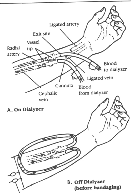
Types of venous access for hemodialysis:
External shunt.
Cannula is placed in a large vein and a large artery that approximate each other.
External shunts, which provide easy and painless access to bloodstream, are prone to infection and clotting and cause erosion of the skin around the insertion area.
Daily cleansing and application of a sterile dressing.
Prevention of physical trauma and avoidance of some activities, such as swimming.
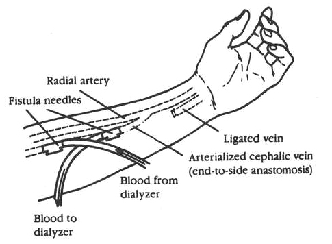
Arteriovenous fistulas or graft.
Large artery and vein are sewn together (anastomosed) below the surface of the skin (fistula) or subcutaneous graft using the saphenous vein, synthetic prosthesis, or bovine xenograft to connect artery and vein.
Purpose is to create one blood vessel for withdrawing and returning blood.
Advantages: greater activity range than AV shunt and no protective asepsis.
Disadvantage: necessity of two venipunctures with each dialysis.
Vein catheterization.
Femoral or subclavian vein access is immediate.
May be short- or long-term duration.
Complications during hemodialysis:
Dysequilibrium syndrome - rapid removal of urea from blood —> reverse osmosis, with water moving into brain cells —» cerebral edema —> possible headache, nausea, vomiting, confusion, and convulsions; usually occurs with initial dialysis treatments; shorter dialysis time and slower rate minimizes.
Hypotension - results from excessive ultrafiltration or excessive antihypertensive medications.
Hypertension - results from volume overload (water and/or sodium), causing dysequilibrium syndrome or anxiety.
Transfusion reactions.
Arrhythmias -due to hypotension, fluid overload, or rapid removal of potassium.
Psychologicalproblems:
Patients react in varying ways to dependence on hemodialysis.
Nurse needs to identify patient reactions and defense mechanisms and to employ supportive behaviors, e.g., include patient in care; continual repetition and reinforcement; do not interpret patient’s behavior—for example, do not say, “You’re being hostile” or “You’re acting like a child”; answer questions honestly regarding quality and length of life with dialysis and/or transplantation; encourage independence as much as possible.
Intermittent peritoneal dialysis: involves introduction of a dialysate solution into the abdomen, where the peritoneum acts as the semipermeable membrane between the solution and blood in abdominal vessels. Procedure:
Area around umbilicus is prepared and anesthetized with local anesthetic, and a catheter is inserted into the peritoneal cavity through a trocar; the catheter is then sutured into place to prevent displacement.
Warmed dialysate is then allowed to flow into the peritoneal cavity. Inflow time: 5-10 min; , Mg!t, K+, Cl', and lactate or acetate.
When solution bottle is empty, dwell time (exchange time) begins. Dwell time: 10-20 min; processes of diffusion, osmosis, and filtration begin to move waste products from bloodstream into peritoneal cavity.
Draining of the dialysate begins with the unclamping of the outflow clamp. Outflow time: usually 10 min; returns less than 2 liters usually result from incomplete peritoneal emptying; turn side to side to increase return; multiple exchanges in 24 hr depending on patient need.
Continuous ambulatory peritoneal dialysis (CAPD): functions on the same principles as peritoneal dialysis, yet allows greater freedom and independence for dialysis patients. Procedure:
Dialysis solution is infused into peritoneum three times daily and once before bedtime.
Dwell time - 24 hour for each daily exchange, and overnight for the fourth (8-12 hours).
Indwelling peritoneal catheter is connected to solution bag at all times - serves to fill and drain peritoneum; concealed in cloth pouch, strapped to the body during dwell time; patient can move about doing usual activities.
Continuous cycling peritoneal dialysis: same principles as CAPD, except uses an automated system to infuse and remove dialysate; reduces nursing care needs with hospitalized patients. Cumbersome equipment inhibits nighttime mobility. Procedure:
Long exchanges without an automated cycler during the day and short exchanges with a cycler at night.
Dwell time - 6-8 hr while sleeping.
Automatic alarms to prevent malfunction with home use.
Kidney transplantation: placement of a donor kidney (from sibling, parent, cadaver) into the iliac fossa of a recipient and the anastomosis of its ureter to the bladder of the recipient; indicated in end-stage renal disease.
Criteria for recipient: irreversible kidney function; under 70 yr of age; patent and functional lower urinary tract; and good surgical risk, free of serious cardiovascular complications. Contraindicated in those with another life-threatening condition.
Donor selection:
Sibling or parent - survival rate of kidney is greater; preferred for transplantation.
Cadave - greater rate of rejection following transplantation, although majority of transplantations are with cadaver kidneys.
Bilateral nephrectomy: necessary for patients with rapidly progressive glomerulonephritis, malignant hypertension, or chronic kidney infections; prevents complications in transplanted kidney.
Analysis/nursing diagnosis:
Altered urinary elimination related to kidney failure.
Fear related to potential transplant rejection.
Risk for infection related to immunosuppression.
Body image disturbance related to immunosuppression.
Nursing care plan/implementation:
Preoperative:
Goal: promote physical and emotional adjustment.
Informed consent.
Lab work completed - histocompatibility, CBC, urinalysis, blood type and crossmatch.
Skin preparation.
Goal: encourage expression of feelings: origin of donor, fear of complications, rejection.
Goal: minimize risk of organ rejection: give medications: begin immunosuppression (azathioprine, corticosteroids, cyclosporine); antibiotics if ordered.
Goal: health teaching.
Nature of surgery; placement of kidney.
Postoperative expectations: deep breathing, coughing, turning, early ambulation; reverse isolation.
Goal: promote uncomplicated recovery of recipient.
Vital signs; CVP; I&O - urine output usually immediate with living donor; with cadaver kidney may not work for a week or more and dialysis will be needed within 24-48 hr. Report <100 mL/hr immediately.
Isolation: strict reverse isolation with immunosuppression; wear face mask when out of room.
Position: back to nonoperative side; semi-Fowler’s to promote gas exchange.
Indwelling catheter care: strict asepsis; characteristics of urine - report gross hematuria, heavy sediment; clots; perineal care; bladder spasms may occur after removal of catheter.
Activity: ambulate 24 hr after surgery; avoid prolonged sitting.
Weigh daily.
Medications: immunosuppressives; analgesics as ordered (pain decreases significantly after 24 hr).
Drains: irrigate only on physician order; meticulous catheter care.
Diet: regular after return of bowel sounds; liberal amounts of protein; restrict fluids, sodium, potassium only if oliguric.
Goal: observe for signs of rejection - most dangerous complication. Three classifications:
Hyperacute - occurs within 5-10 min up to 48 hr after transplantation (rare).
Acute - most common 7-14 days; varies depending on living (1 wk6 mo) or cadaver ( 1 wk-2 yr) donor.
Azathioprine - an antimetabolite that interferes with cellular division. Side effects:
Gastrointestinal bleeding (give PO form with food).
Bone marrow depression; leukopenia; anemia.
Development of malignant neoplasms.
Infection.
Liver damage.
Prednisone - believed to affect lymphocyte production by inhibiting nucleic acid synthesis: anti-inflammatory action helps prevent tissue damage if rejection occurs. Side effects:
Stress ulcer with bleeding (give with food).
Decreased glucose tolerance (hyperglycemia).
Muscle weakness.
Osteoporosis.
Moon facies.
Acne and striae.
Depression and hallucinations.
Cyclosporine (Neoral, Sandimmune) - polypeptide antibiotic used to prevent rejection of kidney, liver, or heart allografts; PO dose given with room temperature chocolate milk or orange juice in a glass dispenser. Side effects:
Nephrotoxicity (increased BUN, creatinine).
Hypertension.
Tremor.
Hirsutism, gingival hyperplasia.
GI - nausea, vomiting, anorexia, diarrhea, abdominal pain.
No signs of rejection (e.g., no weight gain, oliguria).
No depression.
Patient resumes role responsibilities.
Nephrectomy (radical or partial): removal of kidney through flank, retroperitoneal, abdominal, thoracic, or thoracic-abdominal approach; indicated with malignant tumors, severe trauma, or under certain conditions before renal transplantation.
Analysis/nursing diagnosis:
Pain related to surgical incision.
Risk for infection related to wound contamination.
Risk for aspiration related to vomiting.
Constipation related to paralytic ileus.
Anxiety related to possible loss of function in remaining kidney.
Dysfunctional grieving related to perceived loss.
Nursing care plan/implementation:
Preoperative: Goal: optimize physical and psychological functioning.
Postoperative: Goal: promote comfort and prevent complications.
Fluid balance: daily weight - maintain within 2% of preoperative level.
Evaluation/outcome criteria:
No complications (e.g., hemorrhage, paralytic ileus, wound infection).
Acceptance of loss of kidney.
Renal calculi (urolithiasis): formation of calculi (stones) in renal calyces or pelvis that pass to lower regions of urinary tract - ureters, bladder, or urethra; occurs after age 30, with greatest incidence in men, particularly over age 50.
Pathophysiology: organic crystals form (most consist of calcium salts or magnesium - ammonium phosphate (struvite) —> obstruction, infection; increased backward pressure in kidney —> hydronephrosis —> atrophy, fibrosis of renal tubules.
Risk factors (in over 50% of cases, cause is idiopathic):
Urinary tract infection.
Urinary stasis - obstruction.
Metabolic factors - excessive intake of vitamin D or C, calcium carbonate.
Assessment (depends on size, shape, location of stone):
Subjective data:
Pain: occasional, dull, in loin or back when stones are in calyces or renal pelvis; excruciating in flank area (renal colic), radiating to groin when stones are ureteral.
Nausea associated with pain.
Objective data:
Pallor, sweating, syncope, shock, and vomiting due to pain.
Palpable kidney mass with hydronephrosis.
Fever and pyuria with infection.
Lab data:
Urinalysis: abnormal - pH (acidic or alkaline); RBCs (injury); WBCs (infection); increased - specific gravity; casts; crystals; other organic substances, depending on type of stone (i.e., calcium, struvite, uric acid or cystine); positive culture.
Acid-ash diet with calcium oxalate and calcium phosphate stones, magnesium and ammonium phosphate (struvite) stones.
Alkaline-ash diet with uric acid and cystine stones.
Signs of urinary infection: dysuria, frequency, hematuria; seek immediate treatment.
Prepare for removal if indicated; percutaneous or extracorporeal lithotripsy done for stones < 1 cm; ureterolithotomy may also be ordered for larger stones; nephrectomy in extreme cases.
Evaluation/outcome criteria:
Relief from pain.
No signs of urinary obstruction (e.g., increased flank pain, decreased urine output).
No recurrence of lithiasis (adheres to diet and fluid regimen).
Lithotripsy. Extracorporeal shock wave - a noninvasive mechanical procedure used to break up renal calculi so they can pass spontaneously, in most cases. The trunk of the patient is submerged in distilled water. In addition to being strapped to a frame, the patient may also receive sedation and analgesia for pain from sound waves. The procedure takes 30-45 min, and remaining still is important. An underwater electrode generates shock waves that fragment the stone so it can be excreted in the urine a few days after the procedure. A degree of renal colic O may occur, requiring narcotics for up to 3 days. Nursing measures should encourage ambulation and promote diuresis through forcing fluids. Percutaneous lithotripsy - nephrostomy tract above kidney region; nephroscope used to retrieve calculi. Urinary drainage from incision for 3-4 days is normal. May be required for large fragments remaining after extracorporeal lithotripsy. Nursing measures include: dressing changes to prevent infection and prevent skin breakdown, and administration of antibiotics and narcotics for pain.
Benign prostatic hyperplasia (BPH): bladder outlet obstruction resulting from an enlargement of the prostate gland.
Pathophysiology: prostate enlarges, bulges upward, blocks flow of urine from bladder into urethra —> obstruction —> hydroureter, hydronephrosis.
Risk factors:
Changes in estrogen and androgen levels.
Men >50.
Assessment:
Subjective data - urination:
Difficulty starting stream.
Smaller, less forceful.
Dribbling.
Frequency.
Urgency.
Nocturia.
Retention (incomplete emptying).
Inability to void after ingestion of alcohol or exposure to cold.
Objective data:
Catheterization for residual urine: 25-50 mL after voiding.
Enlarged prostate on rectal exam.
Lab data:
Urine - increased RBC, WBC.
Blood - increased creatinine, PSA.
Analysis/nursing diagnosis:
Urinary retention related to incomplete emptying.
Altered urinary elimination related to obstruction.
Urinary incontinence related to urgency, pressure.
Anxiety related to potential surgery.
Body image disturbance related to threat to male identity.
Nursing care plan/implementation:
Goal: relieve urinary retention.
Catheterization: release maximum of 1000 mL initially; avoid bladder decompression, which results in hypotension, bladder spasms, ruptured blood vessels in bladder; empty 200 mL every 5 min.
Patency: irrigate intermittently or continually, as ordered.
Fluids: minimum 2000 mL/24 hr.
Goal: health teaching.
Preparation for surgery (cystostomy, prostatectomy):
Expectations - indwelling catheter (will feel urge to void).
Avoid pulling on catheter (this increases bleeding and clots).
Bladder spasms common 24 - 48 hr after surgery, particularly with transurethral resection and suprapubic approaches.
Threatening nature of procedure (possibility of impotence with perineal prostatectomy).
Prostatectomy: surgical procedure to relieve urinary retention and frequency caused by benign prostatic hyperplasia or cancer of the prostate.
Types
Transurethral resection (TUR) - removal of obstructive prostatic tissue surrounding urethra by an electric wire (resectoscope) introduced through the urethra; hypertrophy may recur, and TUR repeated; little risk of impotence. Laser also being used.
Suprapubic - low midline incision is made directly over the bladder; bladder is opened and large mass of prostatic tissue is removed through incision in urethral mucosa.
Retropubic - removal of hypertrophied prostatic tissue high in pelvic area through a low abdominal incision; bladder is not opened; patient may remain potent.
Perineal - removal of prostatic tissue low in pelvic area is accomplished through an incision made between the scrotum and the rectum; usually results in impotency and incontinence.
Nursing care plan/implementation:
Preoperative: see X. Prostatic hyperplasia.
Postoperative:
Goal: promote optimal bladder function and comfort.
Urinary drainage: sterile closedgravity system - maintain external traction as ordered.
Reinforce purposes, sensations to expect.
Bladder irrigation to control bleeding, keep clots from forming.
Suprapubic catheter care (suprapubic prostatectomy) - closed-gravity drainage system; observe character, amount, flow of drainage.
After removal:
Observe for urinary drainage q4h for 24 hr.
Skin care.
Report excessive drainage to physician.
Dressings: keep dry, clean; reinforce if necessary (may need to change suprapubic dressing if urinary drainage); notify physician of excessive bleeding.
Observe for signs of:
Bladder distention - distinct mound over pubis, slow drip incollecting bottle; irrigate catheter as ordered.
Increased bleeding - expect and report frank bleeding (if venous bleeding, increase traction on catheter); if bright red drainage, and clots (arterial bleeding), may need surgical control; cool, clammy, pale skin and increased pulse rate indicate shock.
Goal: assist in rehabilitation. Emotional support: fears of incontinence, loss of male identity, impotence.
Goal: health teaching.
Expectations: mild incontinence, dribbling for a while (several months) after surgery; need to void as soon as urge is felt; push fluids.
Exercises: perineal (Kegel) 1-2 days after surgery - buttocks are tightened for a count of ten, 20-50 times daily.
Avoid:
Long auto trips, vigorous exercise, heavy lifting (anything heavier than 10 lb), and sexual intercourse for about 3 wk or until medical permission, because they may increase tendency to bleed.
Alcoholic beverages for 1 mo, because this may cause burning on urination; caffeine, because it causes diuresis.
Tub baths, because they increase chance of infection.
Medications: stool softeners or mild cathartics to decrease straining.
Evaluation/outcome criteria:
Relief of symptoms.
No complications (e.g., hemorrhage, impotence).
Urinary diversion. Incontinent - ileal conduit: anastomosis of ureters to a small portion of the ileum; stoma is called urostomy; urine flow is constant; requires external collection device. Continent - Kock pouch, Indiana pouch: segment of small bowel or colon is used to create a pouch; holds urine without leakage; requires self-catheterization.
Indications:
Congenital anomalies of bladder.
Neurogenic bladder.
Mechanical obstruction to urine flow (e.g., bladder cancer).
Chronic progressive pyelonephritis.
Trauma to lower urinary tract.
Analysis/nursing diagnosis:
Altered urinary elimination related to surgical diversion.
Risk for impaired skin integrity related to leakage of urine.
Risk for infection related to contamination of stoma.
Constipation related to absence of peristalsis.
Body image disturbance related to stoma.
Nursing care plan/implementation:
Preoperative: optimal bowel and stoma site preparation.
Diet: low-residue 2 days followed by clear liquids for 24 hr.
Medications:
Neomycin (for bowel sterilization).
Cathartics (GoLYTELY), enemas.
Site selection: appliance faceplate (incontinent diversion) must bond securely; avoid areas of pressure from clothing (waistline); usual site is right or left lower abdominal quadrant.
Postoperative:
Goal: prevent complications and promote comfort.
Observe for signs of:
Paralytic ileus (common complication) - keep NG tube patent.
Stoma necrosis - dusky or cyanotic color (emergency situation).
Skin care: check for leakage around ostomy bag.
Urinary drainage - stents or catheter in stoma; blood in urine in immediate postoperative period; mucus normal.
Goal: health teaching.
Self-care activities:
Peristomal skin care - prevent irritation, breakdown; proper cleansing - soap and water; adhesive remover, if needed.
Appliance application and emptying; pouch opening 2-3 mm larger than stoma; do not remove each day; change appliance every 3-5 days or when leaking.