PROTECTIVE FUNCTIONS

  1. Burns: wounds caused by exposure to excessive heat, chemicals, fire, steam, radiation, or electricity; most often related to carelessness or ignorance; 10,000-12,000 deaths annually; survival best at ages 15 - 30 yr and in burns covering less than 20% of total body surface
    1. Pathophysiology:
      1. Emergent phase (injury to 72 hr): shock due to pain, fright, or terror —> fatigue, failure of vasoconstrictor mechanisms —> hypotension. Capillary dilation, increased permeability —» plasma loss to blisters, edema —> hemoconcentration —» hypovolemia —> hypotension —» decreased renal perfusion —> potential shut-down.
      2. Acute phase (3-5 days): interstitial to plasma fluid shift —> hemodilution —> hypervolemia —» diuresis.
    2. Assessment:
      1. Subjective data: how the burn occurred
      2. Objective data:
        1. Extent of body surface involved: “rule of nines” - head and both upper extremities, 9% each; front and back of trunk, 18% each; lower extremities, 18% each; and perineum, 1%. Requires adjustment for variation in size of head and lower extremities according to age
        2. Location - facial, perineal, and hand and foot bums have potentially more complications because of poor vascularization.
        3. Depth of burn:
          1. First degree (superficial) - epidermal tissue only; not serious unless large areas involve
          2. Second degree (shallow or deep partial thickness) - epidermal and dermal tissue, hospitalization required if more than 10% of body surface involved (major burn).
          3. Third degree (full thickness) - destruction of all skin layers; requires immediate hospitalization; involvement of 10% of body surface considered major burn.
          4. Fourth degree (deep penetrating) - muscles (fascia), bone
        4. Indications of airway burns, singed nasal hair, progressive hoarseness, sooty expectoration; edema may occur in 1 hr; increased mortality rate
        5. Poorer prognosis - infants, due to immature immune system and effects of fluid loss; elderly, due to degenerative diseases and poor healing
        6. Medical history - presence of hypertension, diabetes, alcohol abuse, or chronic obstructive pulmonary disease increases complication rate
    3. Analysis/nursing diagnosis:
      1. Impaired skin integrity related to thermal injury.
      2. Pain (depending on type of burn) related to exposure of sensory receptors.
      3. Fluid volume excess or deficit related to hemodynamic changes.
      4. Risk for infection related to destruction of protective skin.
      5. Impaired gas exchange related to airway injury.
      6. Body image disturbance related to scarring, disfigurement.
      7. Ineffective individual or family coping related to traumatic experience
    4. Nursing care plan/implementation:
      1. Goal: alleviate pain, relieve shock, and maintain fluid and electrolyte balance
        1. Medications: give opioid analgesic and anxiolytics incrementally.
        2. Fluids: IV therapy ; colloids, crystalloids, or 5% dextrose according to burn formula
        3. Monitor hydration status:
          1. Insert indwelling catheter.
          2. Note color, odor, and amount of urine hourly.
          3. Strict intake and output; hourly for 36 hr with large burns.
          4. Check hematocrit (normal: men >40%; women >37%). Increased Het with intravascular fluid depletion.
          5. Weigh daily
      2. Goal: prevent physical complications.
        1. Vital signs: hourly; central venous pressure (CVP) for signs of shock or fluid overload with at-risk patients.
        2. Assess respiratory function (particularly with head, neck burns); patent airway; breath sounds.
        3. Give medications as ordered - tetanus booster; antibiotics to treat documented infection; sedatives and analgesics; antipyretics - avoid aspirin; H 2 blockers.
        4. Isolation: protective; contact isolation (hand washing, protective clothing).
        5. Positioning: turn q2h; prevent contractures.
          1. Head and neck burns - use pillows under shoulders only for hyperextension of neck.
          2. Hand burns - splints.
          3. Arm and hand burns - keep arms at 90-degree angle from body and slightly above shoulders.
          4. Ankle and foot burns - splints; elevate to prevent edema
          5. Splints to maintain functional positions.
          6. ROM exercises according to therapy guidelines; usually several times per day; active exercises most beneficial.
        6. Diet: begin oral fluids at once; food as tolerated - high protein, high calorie for energy and tissue repair (promote positive nitrogen balance); enteral feedings if protein and calorie goals not met.
        7. Observe for: constriction (circumferential or chest wall burns); check peak inspiratory pressure in intubated patient - report increased pressure; check pulses in burned extremities every 1 - 2 hours for 24 hours - report loss of pulses.
      3. Goal: promote emotional adjustment and provide supportive therapy.
        1. Care by same personnel as much as possible, to develop rapport and trust.
        2. Involve patient in care plans.
        3. Answer questions clearly, accurately.
        4. Encourage family involvement and participation.
        5. Provide diversional activities and change furnishings or room adornments when possible, to prevent perceptual deprivation related to immobility.
        6. Point out signs of progress (decreased edema, healing) because patient and family tend to become discouraged and cannot see progress.
        7. Encourage self-care to highest level tolerate
        8. Anticipate psychological changes:
          1. Acute period - severe anxiety: medicate with anxiolytics as ordered; maintain eye contact; explain procedures.
          2. Intermediate period - reactions associated with pain, dependency, depression, anger; give medications to decrease pain; explain procedures; have open, nonjudgmental attitude; use consistent approaches to care; contract with patient regarding division of responsibilities; encourage self-care
          3. Recuperative period - grief process reactivate Anxiety, depression, anger, bargaining, as patient tries to cope with altered body image, leaving security of hospital, finances. Encourage verbalization; refer to support group to assist adaptation.
      4. Goal: promote wound healing - wound care:
        1. Open method - exposure of burns to air; useful in burns of face; thin layer (2-4 mm) topical antimicrobial ointment applied
        2. Closed method - dressings applied to burned areas, changed 1 - 3 times/day; give PO pain medication 30 min before change; IV pain medication during dressing change; tubbing facilitates removal.
        3. Multiple dressing change - common approach; dressings changed bid to q4h depending on wound condition.
        4. Topical antimicrobial therapies.
        5. Tubbing and debridement.
          1. Hydrotherapy - body  temperature bath water; loosens dressings so some float off; soak 20-30 min; encourage limb exercises; do not leave unattended; loss of body heat may occur, with chilling and poor perfusion resulting
          2. Removal of eschar (debridement) - done with forceps and curved scissors; medicate for pain before; use sterile technique; only loose eschar removed, to prevent bleeding; examine wound for infection, color change, decreased granulation - report changes immediately. Chemical debridement also done; agent digests necrotic tissue
        6. Wound coverage, to decrease chances of infection:
          1. Temporary and semipermanent wound coverings.
          2. Autograft - patient donates skin for wound coverage
            1. Types - free grafts (unattached to donor site) and pedicle grafts (attached to donor site).
            2. Procedure - general anesthesia; donor sites shaved and prepared; graft applied to granulation bed; face, hands, and arms grafted first.
            3. Post-skin-graft care:
              1. Sheet grafts: roll cottontipped applicator over graft to remove excess exudate; maintain dressings; aseptic technique; mesh grafts: irrigate as ordered.
              2. Third to fifth day - graft takes on pink appearance if it has taken.
              3. Padding, then splints applied to immobilize grafted extremities.
              4. Pressure garments worn up to 18 months to decrease hypertrophic scarring.
      5. Goal: health teaching.
        1. Mobility needs: exercise; physical therapy; splints, braces.
        2. Community resources: mental health practitioner or psychotherapist if needed for problems with self-image or sexual role; referrals as needed.
        3. Techniques to camouflage appearance: slacks, turtlenecks, long sleeves, wigs, makeup.

      Topical Antimicrobials Used in Burn Care

      Agent Advantages Disadvantages Nursing Implications
      Silver sulfadiazine 1% (Silvadene)

      Broad-spectrum antimicrobial
      Antifungal
      Nonstaining
      Relatively painless
      No systemic metabolic abnormalities

      Less eschar penetration than mafenide acetate (Sulfamylon)
      Decreased granulocyte formation; transient leukopenia
      Macular rash
      Check for allergy to sulfa

      Mafenide acetate
      (Sulfamylon)
      cream or solution

      Eschar penetration
      Effective with Pseudomonas
      Topical of choice for electrical burns Suitable for open or closed method of treatment (cream)
      Used for gram-negative organisms

      Severe pain and burning sensation(lasts 30 min)
      Metabolic acidosis
      Carbonic anhydrase inhibitor
      Ineffective against fungi
      May cause hypersensitivity rash
      Administer pretreatment
      analgesic
      Monitor for metabolic acidosis and hyperventilation Check for allergy to sulfa;
      Check for allergy to sulfa;
      observe for rash
      Silver nitrate Lowcost
      Broad spectrum
      Effective with Candida
      Continuous wet soaks
      Superficial penetration
      Black staining of sheets
      Stinging
      Electrolyte imbalances (low sodium, low chloride, low calcium, low potassium), alkalosis
      Check serum electrolytes daily
      Rewet dressing to keep moist
      Petroleum and mineral oil-based (e.g.. Bacitracin, Neosporin, Polysporin) Bactericidal, gram-positive and gram-negative organisms Painless
      Prevents drying of wound
      Limited ability to penetrate eschar Apply in thin layer (1 mm) Reapply as needed.


      Wound Coverings

      Example Advantages Disadvantages Nursing Implications
      Biological
      Xenograph
      Promotes healing of clean wound; relieves pain; readily available; reduces water and heat loss Easily digested by wound collagenase; allergic reactions Overlap edges slightly; trim away when skin underneath has healed
      Homograft-cadaver skin Reduces water and heat loss; relieves pain; used with antimicrobial mesh; may be left in place until rejection occurs May harbor disease Observe for signs of infection
      Amnion Relieves pain; reduces water and heat loss; has bacteriostatic properties Limited shelf life; requires special preparation for use; may harbor diseases Change cover dressing q48h; leave on wound until it sloughs
      Biosynthetic (Temporary) Biobrane Protects from microbial penetration; decreases pain; promotes healing in partial-thickness burn Not effective for preparing a granulation bed Must be secured to skin with sutures, closure straps, tape, or staples; wrap with gauze; after 48 hr, check for adherence; once adherence has occurred, may be left open to air; check for signs of infection
      Biosynthetic (Semipermanent)Integra Artificial Skin Postexcisional treatment of lifethreatening full or deep partialthickness burns when autograft not available Infection rate lower than autograft Maintain integrity of covering; 18-20 days before thin autograft applied. Avoid hydrotherapy; observe for infection.

    5. Evaluation/outcome criteria:
      1. Return of vital signs to preburn levels.
      2. Minimal to no hypertrophic scarring.
      3. Free of infection; demonstrates wound care.
      4. Maintains functional mobility of limbs; no contractures.
      5. Adjusts to changes in body image; no depression.
      6. Regains independence; returns to work, social activities.
  1. Lyme disease: a spirochetal illness (syndrome); most common tick-borne infectious disease in United States; prevalent Northeast, upper Midwest, and coastal northern California. Reporting is mandatory. With early treatment, recovery is usually quick and complete.
    1. Stages:
      1. Stage I. Rash (erythema migrans) at site ot tick bite; bull’s-eye or target pattern; may appear as hives or cellulitis; common in moist areas (groin, armpit, behind knees). Flulike symptoms may occur (joint pain, chills, fever).
      2. Stage II. If untreated, may progress to cardiac problems (10% of patients) or neurological disturbances - Bell’s palsy (10% of patients); occasionally meningitis, encephalitis, and eye damage may result.
      3. Stage III. From 4 wk to 1 yr after the tick bite, “arthritis,” primarily large joint, develops in half the patients. If untreated, chronic neurological problems may develop.
    2. Assessment (depends on stage): History is important - where do they live or work? Recent travel? Outdoor activities (gardening, hiking, camping, clearing brush)? Knowledge of tick bite and how removed? Pets?
      1. Subjective data:
        1. Malaise (stage I).
        2. Headache (stage I).
        3. Joint, neck, or back pain ( stages I and III).
        4. Weakness (stages II and III).
        5. Chest pain (stage II).
        6. Light-headedness (stage II).
        7. Numbness, pain in arms or legs (stage III).
      2. Objective data:
        1. Rash - erythema migrans (stage I); at least 5 cm/lesion.
        2. Dysrhythmias; heart block (stage II).
        3. Facial paralysis (stage II).
        4. Conjunctivitis, iritis, optic neuritis (stage
        5. Lab data: Lyme titer - elevated (stages II and III). Often inconclusive.
        6. Diagnostic tests: isolation of Borrelia burgdorferi in tissue or body fluid; diagnostic levels of IgM or IgG antibodies in serum or CSF.
    3. Analysis/nursing diagnosis:
      1. Anxiety related to diagnosis.
      2. Pain related to joint inflammation.
      3. Fatigue related to viral illness.
      4. Impaired physical mobility related to joint pain.
      5. Altered thought processes related to neurological deficit.
      6. Decreased cardiac output related to dysrhythmias.
      7. Knowledge deficit (learning need) related to treatment and course of disease.
    4. Nursing care plan/implementation:
      1. Goal: minimize irreversible tissue damage and complications.
        1. Medications according to presenting symptoms: stage I - oral antibiotics for 21 days (doxycycline, amoxicillin, cefotaxime); stages II and III - oral (see stage I) or intravenous antibiotics for 21-28 days (ceftriaxone).
        2. If hospitalized, monitor vital signs q4h for increased temperature, signs of heart failure; check level of consciousness and cranial nerve functioning.
        3. Note treatment response: worsening of symptoms during first 24 hr; redder rash, higher fever, greater pain (Jarisch-Herxheimer reaction).
      2. Goal: alleviate pain, promote comfort.
        1. Medications: salicylates, nonsteroidal anti-inflammatory agents, or other analgesic, as ordered; observe for side effects (GI irritation).
        2. Rest: give instructions on relaxation techniques; create a quiet environment.
        3. Goal: maintain physical and psychological well-being.
          1. Activity: ROM at regular intervals; medicate for pain before exercise; encourage proper posture to reduce joint stress; rest periods between activities and treatments.
          2. Referral: occupational or physical therapy as appropriate.
          3. Reassurance: give psychological support; encourage discussion of feelings.
        4. Goal: health teaching.
          1. Information on disease. Transmission from tick not likely if removed before 48 hours of attachment.
          2. Instructions for home IV antibiotics with heparin lock, if ordered.
          3. Side effects of antibiotics (drug specific); importance of completing therapy.
          4. Signs of disease recurrence (later stages of disease: less severe attacks).
          5. Preventing subsequent infections: wear proper clothing and tick repellent on clothing (20-30% DEET); conduct “tick checks” of self, children, and pets; proper tick removal (use tweezers, steady, gentle traction).
          6. Start vaccination series (LYMErix); three injections at 0, 1, and 12 months.
    5. Evaluation/outcome criteria:
      1. Achieves reasonable comfort.
      2. Regains normal physiological and psychological functioning - no irreversible complications; vital signs within normal limits.
      3. Resumes previous activity level; returns to work
      4. Adheres to follow-up care recommendations.
      5. Knows ways to minimize risk of reinfection.
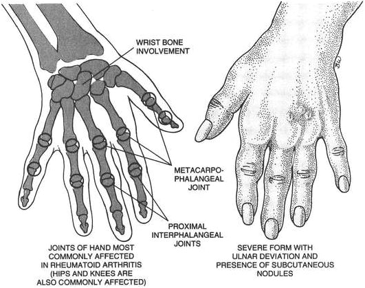
  1. Rheumatoid arthritis: chronic, systemic, collagen, inflammatory disease; etiology unknown; may be autoimmune, viral, or genetic; affects primarily women age 20 - 40 yr; present in 2-3% of total population; follows a course of exacerbations and remissions.
    1. Pathophysiology: synovitis with edema —> proliferation of various blood material (formation of pannus) —> destruction and fibrosis of cartilage (fibrous ankylosis); calcification of fibrous tissue (osseous ankylosis).
    2. Assessment:
      1. Subjective data:
        1. Joints: pain; morning stiffness; swelling.
        2. Easily fatigues; malaise.
        3. Anorexia; weight loss.
      2. Objective data:
        1. Subcutaneous nodules over bony prominences.
        2. Bilateral symmetrical involvement of joints: crepitation, creaking, grating.
        3. Deformities: contractures, muscle atrophy.
        4. Lab data: blood: decreased - hemoglobin/hematocrit; RBCs; increased - WBCs (12,000-15,000), sedimentation rate (>20 mm/hr), rheumatoid factor.
    3. Analysis/nursing diagnosis:
      1. Pain related to joint destruction.
      2. Impaired physical mobility related to joint contractures.
      3. Risk for injury related to the inflammatory process.
      4. Body image disturbance related to joint deformity.
      5. Self-care deficit related to musculoskeletal impairment.
      6. Risk for activity intolerance related to fatigue and stiffness.
      7. Altered nutrition, less than body requirements, related to anorexia and weight loss.
      8. Self-esteem disturbance related to chronic illness.
    4. Nursing care plan/implementation:
      1. Goal: prevent or correct deformities.
        1. Activity:
          1. Bed rest during exacerbations.
          2. Daily ROM - active and passive exercises even in acute phase 5-10 min periods; avoid fatigue and persistent pain.
          3. Heat or pain medication before exercise.
        2. Medications: aspirin (high dosages); nonsteroidals; steroids; antacids given for possible GI upset with ASA, steroids; diseasemodifying antirheumatics (methotrexate, hydroxychloroquine, sulfasalazine).
        3. Fluids: at least 1500 mL liquid daily to avoid renal calculi; milk for GI upset.
      2. Goal: health teaching.
        1. Side effects of medications: tarry stools (GI bleeding); tinnitus (ASA).
        2. Psychosocial aspects: possible need for early retirement; financial hardship; loss of libido; unsatisfactory sexual relations.
        3. Prepare for joint repair or replacement if indicated.
    5. Evaluation/outcome criteria:
      1. Remains as active as possible; limited loss of mobility; performs self-care activities.
      2. No side effects from drug therapy (e.g., GI bleeding).
      3. Copes with necessary lifestyle changes; complies with treatment regimen.

Rheumatoid arthritis.

  1. Lupus erythematosus: chronic inflammatory disease of connective tissue; may affect or involve any organ; vague etiology, but genetic factors, viruses, hormones, and drugs are being investigated; occurs primarily in women ages 18-35 yr. Two forms: discoid lupus erythematosus (DLE) affects skin only, and systemic lupus erythematosus (SLE) affects multiple organs.
    1. Pathophysiology: possible toxic effects from immune complexes deposited in tissue (antibodyantigen trapping in organ capillaries) - fibrinoid necrosis of collagen in connective issue, small arterial walls (kidneys and heart particularly) cellular death, obstructed blood flow.
    2. Assessment:
      1. Subjective data:
        1. Pain: joints.
        2. Anorexia; weight loss.
        3. Photophobia; sensitivity to sun.
        4. Weakness.
        5. Nausea, vomiting.
      2. Objective data:
        1. Fever.
        2. Rash: butterfly distribution across nose, cheeks.
        3. Lesions: raised, red, scaling plaques - coinlike (discoid).
        4. Ulceration: oral or nasopharyngeal.
        5. Lab data:
          1. Blood: increased LE cells; decreased - RBCs, WBCs, thrombocytes.
          2. Urine - hematuria, proteinuria (nephritis).
    3. Analysis/nursing diagnosis:
      1. Risk for injury related to possible autoimmune disorder.
      2. Pain related to joint inflammation.
      3. Risk for activity intolerance related to extreme fatigue, anemia.
      4. Body image disturbance related to chronic skin eruptions.
      5. Altered nutrition, less than body requirements, related to anorexia, nausea, vomiting.
      6. Altered oral mucous membrane related to ulcerations.
    4. Nursing care plan/implementation:
      1. Goal: minimize or limit immune response and complications.
        1. Activity: rest; 8 - 10 hr sleep; unhurried environment; assist with stressful activities; ROM to prevent joint immobility and stiffness.
        2. Skin care: hygiene; topical steroid cream as ordered for inflammation, pruritus, scaling.
        3. Mouth care: several times daily if stomatitis present; soft, bland, or liquid diet to prevent irritation.
        4. Diet: low sodium if edematous; low protein with renal involvement.
        5. Observe for signs of complications:
          1. Cardiac/respiratory (tachycardia, tachypnea, dyspnea, orthopnea).
          2. GI (diarrhea, abdominal pain, distention).
          3. Renal (increased weight, oliguria, decreased specific gravity).
          4. Neurological (ptosis, ataxia).
          5. Hematological (malaise, weakness, chills, epistaxis); report immediately.
        6. Medications, as ordered:
          1. Analgesics.
          2. Anti-inflammatory agents (aspirin, prednisone) and immunosuppressive drugs (azathioprine [Imuran], cyclophosphamide [Cytoxan]) to control inflammation.
          3. Antimalarials for skin and joint manifestations.
      2. Goal: health teaching.
        1. Disease process: diagnosis, prognosis, effects of treatment.
        2. Avoid precipitating factors:
          1. Sun (aggravates skin lesions; thus, cover body as much as possible).
          2. Altering dosage of medications.
          3. Pregnancy requires medical clearance.
          4. Fatigue, stress.
          5. Infections.
        3. Medications: side effects of immunosuppressives and corticosteroids.
        4. Regular exercise: walking, swimming; but avoid fatigue.
        5. Wear Medic Alert bracelet.
    5. Evaluation/outcome criteria:
      1. Attains a state of remission.
      2. No organ involvement (e.g., cardiac, renal complications).
      3. Keeps active within limitations.
      4. Continues follow-up medical care - recognizes symptoms requiring immediate attention.
  1. Acquired immunodeficiency syndrome (AIDS): the terminal stage of the disease continuum caused by human immunodeficiency virus (HIV), a retrovirus; typically progresses from asymptomatic seronegative status to asymptomatic seropositive status to subclinical immune deficiency to lymphadenopathy (early AIDS) to AIDS-related complex (middle stage with combination of symptoms) to AIDS; hallmarks of HIV infection include opportunistic infections: Pneumocystis carinii pneumonia (PCP); cytomegalovirus (CMV); Mycobacterium tuberculosis; hepatitis B; herpes simplex or zoster; candidiasis; may take 7-10 years before signs and symptoms occur.
    1. High-risk populations:
      1. Men, homosexual or bisexual (71%).
      2. Injection drug users (IDU)/heterosexual (10%).
      3. IDU/homosexual (9%).
      4. Hemophiliacs and multiple transfusion recipients (1%).
      5. Heterosexual (5%).
      6. Undetermined/other (4%).
    2. Pathophysiology : abnormal response to foreign antigen stimulation (acquired immunity) —> deficiency in cell-mediated immunity - T lymphocytes, specifically helper cells (T4 cells) and hyperactivity of the humoral system (B cells).
    3. Assessment:
      1. Subjective data:
        1. Fatigue: prolonged; associated with headache or light-headedness.
        2. Unexplained weight loss: >10%.
      2. Objective data:
        1. Fever: prolonged or night sweats >2 weeks
        2. Lymphadenopathy.
        3. Skin or mucous membrane lesions: purplish-red, nodules (Kaposi’s sarcoma).
        4. Cough: persistent, heavy, dry.
        5. Diarrhea: persistent.
        6. Tongue/mouth “thrush”; oral hairy leukoplakia.
        7. Diagnostic tests (with permission of patient): enzyme-linked immunosorbentassay (ELISA); Western blot test.
        8. Lab data: decreased - CD4 (T4) lymphocytes, hematocrit, WBC, platelets. Seropositive - syphilis, hepatitis B; ELISA - positive; Western blot test - positive (mean time for seroconversion is 6 weeks after infection).
    4. Analysis/nursing diagnosis:
      1. Risk for infection related to immunocompromised state.
      2. Fatigue related to anemia.
      3. Altered nutrition, less than body requirements, related to anorexia.
      4. Impaired skin integrity related to nonhealing viral lesions, Kaposi’s sarcoma.
      5. Diarrhea related to infection or parasites.
      6. Risk for activity intolerance related to shortness of breath.
      7. Ineffective airway clearance related to pneumonia.
      8. Visual sensory/perception alteration related to retinitis.
      9. Risk for altered body temperature (fever) related to opportunistic infections.
      10. Social isolation related to stigma attached to AIDS.
      11. Powerlessness related to inability to control disease progression.
      12. Altered thought processes related to dementia.
      13. Ineffective individual coping related to poor prognosis.
      14. Risk for violence, self-directed, related to anger, panic, or depression.
    5. Nursing care plan/implementation:
      1. Goal: reduce risk of infection; slow disease progression.
        1. Observe signs of opportunistic infections: weight loss, diarrhea, skin lesions, sore throat.
        2. Monitor vital signs (including temperature).
        3. Note secretions and excretions: changes in color, consistency, or odor indicating infection.
        4. Diet: monitor fluid and electrolytes; strict measurement; encourage adequate dietary intake (high calorie, high protein, low bulk); 5-10 times RDA for watersoluble vitamins (B complex, C); favorite foods from home; enteral feedings. Six small meals/day.
        5. Protective isolation, if indicated, for severe immunocompromise.
        6. Antiviral medications, as ordered: nucleoside reverse transcriptase inhibitors, e.g., zidovudine (Retrovir); nonnucleoside reverse transcriptase inhibitors, e.g., nevirapine (Viramune); protease inhibitors, e.g., indinavir sulfate (Crixivan); drug toxicity and numerous side effects likely (rash, GI upset); large number of pills and tight administration schedule; costly; potential for drug resistance.
      2. Goal: prevent the spread of disease.
        1. Frequent hand washing, even after wearing gloves.
        2. Avoid exposure to blood, body fluids of patient; wear gloves, gowns; proper disposal of needles, IV catheters.
      3. Goal: provide physical and psychological support.
        1. Oral care: frequent.
        2. Cooling bath: 1:10 concentration of isopropyl alcohol in tepid water; avoid plastic-backed pads with night sweats.
        3. Encourage verbalization of fears, concerns without condemnation; may suffer loss of job, lifestyle, significant other.
        4. Determine status of support network: arrange contact with support group.
        5. Observe for severe emotional symptoms (suicidal tendencies).
        6. Address issues surrounding death to ensure quality of life: advance directive prepared and on file; code blue status; reassurance of comfort and pain control.
      4. Goal: health teaching.
        1. Avoidance of environmental sources of infection (kitty litter, bird cages, tub bathing).
        2. Precautions following discharge: riskreducing behaviors; condoms (latex), limit number of sexual partners, avoid exposure to blood or semen during intercourse.
        3. Family counseling; availability of community resources.
        4. Information on disease progression and life span.
        5. Stress-reduction techniques: visualization, guided imagery, meditation.
        6. Expected side effects with drug therapy; importance of compliance.
    6. Evaluation/outcome criteria:
      1. Relief of symptoms (e.g., afebrile, gains weight).
      2. Resumes self-care activities; returns to work; improved quality of life.
      3. Accepts diagnosis; participates in support group.
      4. Progression of disease slows; improved survival probability.
      5. Retains autonomy, self-worth.
      6. Permitted to die with dignity.
TABLE 7-21 Standard Precautions