Imbalances in fluid and electrolytes may be due to changes in the total quantity of either substance (deficit or excess), protein deficiencies, or extracellular fluid volume shifts. Older patients and very young patients are particularly susceptible.

  1. Fluid volume deficit (dehydration): mechanism that influences fluid balance and sodium levels; decreased quantities of fluid and electrolytes may be caused by deficient intake (poor dietary habits, anorexia, and nausea), excessive output (vomiting, nasogastric suction, and prolonged diarrhea), or failure of regulatory mechanism that influences fluid balance and sodium levels.
    1. Pathophysiology: water moves out of the cells to replace a significant water loss; cells eventually become unable to compensate for the lost fluid, and cellular dehydration begins, leading to circulatory collapse.
    2. Risk factors:
      1. No fluids available.
      2. Available fluids not drinkable.
      3. Inability to take fluids independently.
      4. No response to thirst; does not recognize the need for fluids.
      5. Inability to communicate need; does not speak same language.
      6. Aphasia.
      7. Weakness, comatose.
      8. Inability to swallow.
      9. Psychological alterations.
      10. Overuse of diuretics.
      11. Increased vomiting.
      12. Fever.
      13. Wounds, burns.
      14. Blood loss.
    3. Assessment:
      1. Subjective data
        1. Thirst.
        2. Behavioral changes: apprehension, apathy, lethargy, confusion, restlessness.
        3. Dizziness.
        4. Numbness and tingling of hands and feet.
        5. Anorexia and nausea.
        6. Abdominal cramps.
      2. Objective data
        1. Sudden weight loss of 5%.
        2. Vital signs:
          1. Decreased BP; postural changes.
          2. Increased temperature.
          3. Irregular, weak, rapid pulse.
          4. Increased rate and depth of respirations.
        3. Skin: cool and pale in absence of infection; decreased turgor.
        4. Urine: oliguria to anuria, high specific gravity.
        5. Eyes: soft, sunken.
        6. Tongue: furrows.
        7. Lab data:
          1. Blood - increased hematocrit and BUN.
          2. Urine - decreased 17-ketosteroids.
    4. Analysis/nursing diagnosis:
      1. Fluid volume deficit related to inadequate fluid intake.
    5. Nursing care plan/implementation:
      1. Goal: restore fluid and electrolyte balance - increase fluid intake to hydrate client.
        1. IVs as ordered; small, frequent drinks by mouth.
        2. Daily weights (same time of day) to monitor progress of fluid replacement.
        3. I&O, hourly outputs (when in acute state).
        4. Avoid hypertonic solutions (may cause fluid shift when compensatory mechanisms begin to function).
      2. Goal: promote comfort.
        1. Frequent skin care (lack of hydration causes dry skin, which may increase risk
      3. Decreased or inadequate output of urine.
      4. Psychogenic polydipsia.
      5. Certain medical conditions: tuberculosis; encephalitis; meningitis; endocrine disturbances; tumors of lung, pancreas, duodenum, heart failure.
      6. Inadequate kidney function or kidney failure.
    1. Assessment:
      1. Subjective data
        1. Behavioral changes: irritability, apathy, confusion, disorientation.
        2. Headache.
        3. Anorexia, nausea, cramping.
        4. Fatigue.
        5. Dyspnea.
      2. Objective data
        1. Vital signs: elevated blood pressure.
        2. Skin: warm, moist; edema - eyelids, facial, dependent, pitting.
        3. Sudden weight gain of 5 lbs.
        4. Pink, frothy sputum; productive.
        5. Constant, irritating cough.
        6. Crackles in lungs.
        7. Pulse, bounding.
        8. Engorgement of neck veins in sitting position.
        9. Urine: polyuria, nocturia.
        10. Lab data:
          1. Blood - decreasing hematocrit, BUN.
          2. Urine - decreasing specific gravity.
    2. Analysis/nursing diagnosis:
      1. Fluid volume excess related to excessive fluid intake or decreased fluid output.
    3. Nursing care plan/implementation:
      1. Goal: maintain oxygen to all cells.
        1. Position: semi-Fowler’s or Fowler’s to facilitate improved gas exchange.
        2. Vital signs: q4h.
        3. Fluid restriction.
        4. Possible rotating tourniquets as needed (especially for interstitial-to-plasma shift).
      2. Goal: promote excretion of excess fluid.
        1. Medications as ordered: diuretics.
        2. If in kidney failure: may need dialysis; explain procedure.
        3. Assist patient during paracentesis, thoracentesis, phlebotomy.
          1. Monitor vital signs to detect shock.
          2. Prevent injury by monitoring sterile technique.
          3. Prevent falling by stabilizing appropriate position during procedure.
          4. Support patient psychologically.
      3. Goal: obtain/maintain fluid balance.
        1. Daily weights; 1 kg = 1000 mL fluid.
        2. Measure: all edematous parts, abdominal girth, I&O.
        3. Limit: fluids by mouth, IVs, sodium.
        4. Strict monitoring of IV fluids.
      4. Goal: prevent tissue injury.
        1. Skin and mouth care as needed.
        2. Evaluate feet for edema and discoloration when patient is out of bed.
        3. Observe suture line on surgical patients (potential for evisceration due to excess fluid retention).
        4. IV route preferred for parenteral medications; Z track if medications are to be given IM (otherwise injected liquid will escape through injection site).
      5. Goal: health teaching.
        1. Improve nutritional status with lowsodium diet.
        2. Identify cause that put patient at risk for imbalance, methods to avoid this situation in the future.
        3. Desired and side effects of diuretics and other prescribed medications.
        4. Monitor urinary output, ankle edema; report to health care manager when fluid retention is noticed.
        5. Limit fluid intake when kidney/cardiac function impaired.
    4. Evaluation/outcome criteria:
      1. Fluid balance obtained.
      2. No respiratory, cardiac complications.
      3. Vital signs within normal limits.
      4. Urinary output improved, no evidence of edema.
  1. Common electrolyte imbalances: electrolytes are taken into the body in foods and fluids; normally lost through sweat and urine. May also be lost through hemorrhage, vomiting, and diarrhea. Electrolytes have major influences on: body water regulation and osmolality, acid-base regulation, enzyme reactions, and neuromuscular activity. Clinically important electrolytes are:
    1. Sodium (Na+): normal 135-145 mEq/L.| Most prevalent cation in extracellular fluid. Controls osmotic pressure; essential for neuromuscular functioning and intracellular chemical reactions. Aids in maintenance of acid-base balance. Necessary for glucose to be transported into cells.
      1. Hyponatremia - sodium deficit, resulting from either a sodium loss or water excess. Serum-sodium level below 135 mEq/L; symptoms usually do not occur until below 120 mEq/L unless rapid drop.
      2. Hypernatremia - excess sodium in the blood, resulting from either high sodium intake, water loss, or low water intake. Serum-sodium level above 145 mEq/L,
    2. Potassium (K+):normal 3.5-5.0 mEq/L Direct effect on excitability of nerves and muscles. Contributes to intracellular osmotic pressure and influences acid-base balance. Major cation of the cell. Required for storage of nitrogen as muscle protein.
      1. Hypokalemia - potassium deficit related to dehydration, starvation, vomiting, diarrhea, diuretics. Serum-potassium level below 3.5 mEq/L; symptoms may not occur until below 2.5 mEq/L.
      2. Hyperkalemia - potassium excess related to severe tissue damage, renal disease, excess administration of oral or IV potassium. Serumpotassium level above 5 mEq/L; symptoms usually occur when above 6.5 mEq/L.
    3. Calcium (Ca2+): normal 4.5-5.5 mEq/L.| Essential to muscle metabolism, cardiac function, and bone health. Controlled by parathyroid hormone; reciprocal relationship between calcium and phosphorus.
      1. Hypokalemia - potassium deficit related to dehydration, starvation, vomiting, diarrhea, diuretics. Serum-potassium level below 3.5 mEq/L; symptoms may not occur until below 2.5 mEq/L.
      2. Hyperkalemia - potassium excess related to severe tissue damage, renal disease, excess administration of oral or IV potassium. Serumpotassium level above 5 mEq/L; symptoms usually occur when above 6.5 mEq/L.
    4. Magnesium (Mg2+): normal 1.5-2.5 mEq/L.| Essential to cellular metabolism of carbohydrates and proteins.
      1. Hypomagnesemia - magnesium deficit related to impaired absorption from GI tract, excessive loss through kidneys, and prolonged periods of poor nutritional intake. Hypomagnesemia leads to neuromuscular irritability. Serum-magnesium level below 1.5 mEq/L.
      2. Hypermagnesemia - magnesium excess related to renal insufficiency, overdose during replacement therapy, severe dehydration, repeated enemas with Mg2+ sulfate (epsom salts). Serum-magnesium level above 2.5 mEq/L.


Electrolyte Imbalances

Disorder and Related Conditions Assessment Analysis/Nursing Diagnosis Nursing Care Plan/ Implementation Evaluation/Outcome Criteria
Subjective Data Objective Data
Hyponatremia
  • Addison’s disease
  • Starvation
  • Gi suction
  • Thiazide diuretics
  • Excess water intake, enemas
  • Fever
  • Fluid shifts
  • Ascites
  • Bums
  • Small-bowel obstruction
  • Profuse perspiration
  • Apathy, apprehension, mental confusion. delirium
  • Fatigue
  • Vertigo, headache
  • Anorexia, nausea
  • Abdominal and
    muscle cramps
  • Pulse: rapid and weak
    BP: postural hypotension
    Shock, coma
  • GI: weight loss, diarrhea, loss
    through NG tubes
  • Muscle weakness
  • Diarrhea
  • Fluid volume excess
  • Altered nutrition, less than
    body requirements
  • Sensory-perceptual alteration (kinesthetic)
  • Obtain normal sodium level:
    identify cause of deficit, increase sodium intake PO (salty
    foods). IVs - hypertonic solu-
    tions
  • Prevent further sodium loss: irrigate NG tubes with saline;
  • hourly I&O to monitor kidney
    output
  • Prevent injury related to shock,
    dizziness, decreased sensorium;
    dangle before ambulation
  • Skin care
  • No complications of shock
  • present
  • Return of muscle strength
  • Alert, oriented
  • Limits intake of plain water
Hypernatremia
  • High sodium intake
  • Low water intake
  • Diarrhea
  • High fever with rapid
    respirations
  • Impaired renal functions
  • Acute tracheobronchitis
  • Lethargy
  • Restlessness, agitation
  • Confusion
  • BP and temperature elevated
    Neuromuscular: diminished reflexes
  • Skin: flushed; firm turgor
  • GI: mucous membrane dry,
    sticky
  • GU: decreased output
  • Fluid volume deficit
  • Fluid volume excess
  • Altered nutrition, less than
    body requirements
  • Sensory-perceptual alteration
  • Obtain normal sodium level: de-
    w crease sodium intake
  • I&O to recognize signs and symp-
    toms of complications, e.g.,
    heart failure, pulmonary edema
  • No complaint of thirst
  • Alert, oriented
  • Relaxed in appearance
  • Identifies high-sodium foods to avoid
Hypokalemia
  • Decreased intake:
  • Poor potassium food intake
  • Excessive dieting
  • Nausea
  • Alcoholism
  • IV fluids without added
  • potassium
  • Increased loss:
  • GI suctioning, vomiting, diarrhea
  • Ulcerative colitis
  • Drainage: ostomy, fistulas
  • Medications: potassium-losing diuretics,digoxin, cathartics
  • Increased aldosterone production
  • Renal disorders
  • Apathy, lethargy, fatigue, weakness
  • Irritability, mental
    confusion
  • Anorexia, nausea
    Leg cramps
  • Muscles: weakness, paralysis,
    paresthesia, hyporeflexia
  • Respirations: shallow to respiratory arrest
  • Cardiac decreased BP; elevated, weak, irregular pulse;
    arrhythmias
  • ECG: low, flat T waves; prolonged ST segment: elevated
    U wave; potential arrest
  • GI: vomiting, flatulence, constipation; decreased motility
    - distention - paralytic
    ileus
  • GU: urine not concentrated;
    polyuria, nocturia; kidney
    damage
  • Speech: slow
  • Decreased cardiac output
  • Fatigue
  • Altered cardiopulmonary tissue
  • perfusion
  • Ineffective breathing patterns
    Constipation
    Bathing/hygiene self-care
    deficit
  • Impaired home maintenance
    management
  • Sensory-perceptual alteration (gustatory)
  • Replace lost potassium: increase potassium in diet (see Unit 9); liquid PO potassium medications dilute in juice to aid
    taste; give potassium only if
    kidneys functioning
  • Prevent injury to tissues: prevent
    infiltration, pain, tissue damage
  • Prevent potassium loss: irrigate NG
    tubes with saline, not water
  • Identifies cause of imbal-
    ance
  • Lists foods to include in diet
  • Lists signs and symptoms of
    imbalance
  • Return of muscle strength
  • No cardiac arrhythmias
Hyperkalemia
  • Burns
  • Crushing injuries
  • Kidney disease
  • Excessive infusion or ingestion of K *
  • Adrenal insufficiency
  • Mercurial poisoning
  • Irritability
  • Weakness, muscle cramps
  • Nausea, intestinal cramps
  • Musc/ex paresthesia, flaccid
    muscle paralysis (later)
  • Cardiac: irregular pulse; arrhythmias; bradycardia -
    asystole
  • ECG: high T waves; depressed
    ST segment, widened QRS
    complex; diminished or absent P waves; ventricular fibrillation
  • GI: explosive diarrhea; hyper-
    active bowel sounds
  • Kidney: scanty to no urine
  • Decreased cardiac output
  • Altered urinary elimination
  • Activity intolerance
  • Ineffective breathing patterns
  • Diarrhea
  • Impaired home maintenance management
  • Decrease amount of potassium in
    body; identify
    and treat cause of
    imbalance; give
    foods low in K";
    avoid
    drugs or IV fluids containing K*
  • If kidney failure present, may
    need to prepare for dialysis
  • [K- 3^5.0 rntyr ]
  • No complications (e.g., arrhythmias, acidosis, respiratory failure)
Hypocalcemia
  • Acute pancreatitis
  • Diarrhea
  • Peritonitis
  • Damage to parathyroid
    during thyroidectomy
  • Hypothyroidism
  • Bums
  • Pregnancy and lactation
  • Iow vitamin D intake
  • Multiple blood transfusions
  • Renal disorders
  • Massive infection
  • Fatigue
  • Tingling/numbness;
    fingers and circumoral
  • Abdominal cramps
  • Palpitations
  • Dyspnea
  • Muscle spasms: tonic muscles,
    carpopedal, laryngeal
  • Neuromuscular: grimacing, hy-
    perirritable facial nerves
  • Tetany - convulsions
  • Orthopedic: osteoporosis -
    fractures
  • Cardiac: arrhythmias - arrest
    GL diarrhea
  • Pain
  • Diarrhea
  • Altered nutrition, less than
    body requirements
  • Risk for injury
  • Sensory-perceptual alteration (gustatory)
  • Prevent tetany (medical emer-
    40 gency): calcium gluconate IV,
  • 2.5-5.O ml. 10% solution; re-
    peated qlOmin to maximum
    dose of 30 mL
  • Prevent tissue injury due to hy-
    poxia and sloughing; adminis-
    ter slowly;
    avoid infiltration
  • Prevent injury related to medica-
    tion administration. Caution:
    drug interaction with carbon-
    ate, phosphate, digitalis;
    avoid
    hypercalcemia
  • In less acute condition: increase
    ten calcium intake—calcium glu-
    conate or lactate
  • No signs of tetany
  • Absent Trousseau's and
    Chvostek's signs
  • List foods high in vitamin D and calcium
Hypercalcemia
  • Parathyroid glands:
    overactive, tumor
  • Increased immobility
    Decreased renal function
  • Bone cancer
  • Increased vitamin D and
    calcium intake
  • Milk-alkali syndrome -
    self-administration of
    antacids; increased
    milk in diet to relieve
    GI symptoms
  • Pain: flank, deep
    bone, shin splints
  • Muscle weakness, fa-
  • tigue
  • Anorexia, nausea
  • Headache
  • Thirst —» polyuria
  • Muscles: relaxed
  • GU: kidney stones
  • GI: increased milk intake, con-
    stipation. dehydration
  • Neurological: stupor - coma
  • Decreased cardiac output
  • Constipation
  • Activity intolerance
  • Altered urinary elimination
  • Pain
  • Reduce calcium intake: decrease
    m foods high in calcium; identify
    cause of imbalance; give
  • steroids, diuretics as ordered;
    isotonic saline IV
  • Prevent injury: prevent pathological fractures (e.g., advanced
    cancer); prevent renal calculi by
    increasing fluid intake
  • No pain reported
  • No fractures/calculi seen on
    x-ray exam
Hypomagnesemia
  • Impaired GI absorption
    Prolonged malnutrition
    or starvation
  • Alcoholism
  • Excess loss of magnesium through kidneys. related to increased aldosterone
    production
  • Prolonged diarrhea
    Draining Gl fistulas
  • Agitation
  • Depression
  • Confusion
  • Paresthesia
  • Muscles: irritable, tremors,
    spasticity, tetany —» convul-
    sions
  • Cardiac: arrhythmias, tachycardia
 
  • Risk for injury related to
    seizure activity
  • Decreased cardiac output
  • Provide safety: prevent injury to
    disoriented patient; administer
    magnesium salts PO or IV
  • Health teaching: prevention;
  • diet - high-magnesium foods:
    fruits, green vegetables, whole
    grain cereals, milk, meats, nuts
Hypermagnesemia
  • Renal failure
  • Diabetic ketoacidosis
  • Severe dehydration
  • Antacid therapy
  • Drowsiness, lethargy
  • Neuromuscular: loss of deep-
    tendon reflexes
  • Respiratory: depression
  • Cardiac: arrest, hypotension
  • Ineffective breathing pattern
  • Decreased cardiac output
  • Fluid volume deficit
  • Fluid volume excess
  • Altered cardiopulmonary tissue perfusion
  • Obtain norma! magnesium level:
    IV calcium; fluids; possible
    dialysis
  • No complications (e,g„ respiratory depression, arrhythmias)
  • Identifies magnesium-based
    antacids (e.g., Gclusil)
  • Deep-tendon reflexes 2+


Blood Gas Variations with Acid-Base Imbalances
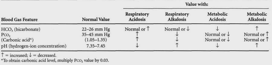


  1. Acid-base balance: concentration of hydrogen ions in extracellular fluid is determined by the ratio of bicarbonate to carbonic acid. The normal ratio is 20:1. Even when arterial blood gases are abnormal, if the ratio remains at 20:1, no imbalance will occur.
    1. Causes of blood gas abnormalities:
    2. Types of acid-base imbalance:
      1. Acidosis: hydrogen ion concentration increases and pH decreases.
      2. Alkalosis: hydrogen ion concentration decreases and pH increases.
      3. Metabolic imbalances: bicarbonate is the problem. In primary conditions, the level of bicarbonate is directly proportional to pH.
        1. Metabolic acidosis: excessive acid is produced or added to the body, bicarbonate is lost, or acid is retained due to poorly functioning kidneys. Deficit of bicarbonate.
        2. Metabolic alkalosis: excessive acid is lost or bicarbonate or alkali is retained. Excess of bicarbonate.
        3. As compensatory mechanism, Pco2 will be low in metabolic acidosis, as the body attempts to eliminate excess carbonic acid and elevate pH. Pco2 will become elevated in metabolic alkalosis.
      4. Respiratory imbalances: carbonic acid is the problem. In primary conditions, Pco2 is inversely proportional to the pH.
        1. Respiratory acidosis: pulmonary ventilation decreases, causing an elevation in the level of carbon dioxide or carbonic acid. Excess of Pco2.
        2. Respiratory alkalosis: pulmonary ventilation increases, causing a decrease in the level of carbon dioxide or carbonic acid. Deficit of Pco2.
        3. As a compensatory mechanism, the level of bicarbonate will increase in respiratory acidosis and decrease in respiratory alkalosis.
    3. Assessment: Acid-Base Imbalances.
    4. Analysis/nursing diagnosis:
      1. Impaired gas exchange related to hyperventilation.
      2. Ineffective breathing pattern related to decreased thoracic movements.
      3. Ineffective airway clearance related to retained secretions.
      4. Risk for injury related to poorly functioning kidneys.
      5. Altered renal tissue perfusion related to dehydration.
      6. Altered urinary elimination, related to renal failure.
      7. Fluid volume excess related to altered kidney function.
      8. Fluid volume deficit related to diarrhea or dehydration.
      9. Knowledge deficit related to self-administration of antacid medications.
    5. Nursing care plan/implementation: Acid-Base Imbalances.
    6. Evaluation/outcome criteria: Acid-Base Imbalances.

Blood Gas Abnormalities: Causes

Decreased Po2

Collapsed alveoli (atelectasis)

  1. Airway obstruction
    1. By the tongue
    2. By a foreign body
  2. Failure to take deep breaths
    1. Pain (rib fracture, pleurisy)
    2. Paralysis of respiratory muscles (spinal cord injury, polio)
    3. Depression of the respiratory center (head injury, drug overdose)
  3. Collapse of the whole lung (pneumothorax)

Fluid in the alveoli

  1. Pulmonary edema
  2. Pneumonia
  3. Near-drowning
  4. Chest trauma

Other gases in the alveoli

  1. Smoke inhalation
  2. Inhalation of toxic chemicals
  3. Carbon monoxide poisoning

Respiratory arrest

Elevated Pco2

Decreased CO2 elimination (hypoventilation)

  1. Decreased tidal volume
    1. Pain (rib fractures, pleurisy)
    2. Weakness (myasthenia gravis)
    3. Paralysis (spinal cord injury, polio)
  2. Decreased respiratory rate
    1. Head injury
    2. Depressant drugs
    3. Stroke

Increased CO2 production

  1. Fever
  2. Muscular exertion
  3. Anaerobic metabolism


Acid-Base Imbalances

Disorder and Related Conditions Assessment Nursing Care Plan/ Implementation Evaluation/Outcome Criteria
Subjective Data Objective Data
Respiratory Acidosis
  • Acute bronchitis
  • Emphysema
  • Respiratory obstruction Atelectasis
  • Damage to respiratory center
  • Pneumonia
  • Asthmatic attack Drug overdose
  • Drug overdose
  • Headache
  • Irritability
  • Disorientation
  • Weakness
  • Dyspnea on exertion
  • Nausea
  • Increased respirations Cyanosis
  • Tachycardia
  • Diaphoresis
  • Dehydration
  • Coma (CO2 narcosis)
  • Hyperventilation to compensate if no pulmonary pathology present
  • IHCO2 normal, Paco, elevated, pH <7.5
  • Assist with normal breathing: encourage coughing;
  • suction airway; postural drainage; pursed-lip
  • B breathing; raise HOB
  • Protect from injury: oxygen at 2 L; encourage fluids; avoid sedation; medica-
  • Health teaching: identify cause, prevent future episodes; increase awareness regarding risk factors and early signs of impending imbalance; encourage compliance
  • Normal acid-base balance obtained
  • Respiratory rate slows, <30
  • No signs of pulmonary infection (e.g., sputum colorless, breath sounds clear)
  • Demonstrates breathing exercises (e.g., diaphragmatic breathing)
Metabolic Acidosis
  • Diabetic ketoacidosis
  • Hyperthyroidism
  • Severe infections
  • Lactic acidosis in shock
  • Renal failure - uremia
  • Prolonged starvation diet; low-protein diet
  • Diarrhea, dehydration
  • Hepatitis
  • Burns
  • Headache
  • Restlessness
  • Apathy
  • weakness
  • Disorientation
  • Thirst
  • Nausea, abdominal pain
  • Kussmaul’s respirations: deep, rapid air hunger;
  • Ttemperature
  • Vomiting, diarrhea
  • Dehydration
  • Stupor - convulsions -
  • HCO2 below normal
  • Paco, normal, K+ >5, pH <7.39
Restore normal metabolism: correct underlying problem; sodium bicarbonate PO/IV; sodium lactate; fluid replacement, Ringer’s solution; Prevent complications: regular insulin for ketoacidosis; hourly outputs; prepare for dialysis if in kidney failure Health teaching: identify signs and symptoms of primary illness; prevent complications, cardiac arrest; diet instructions
  • Normal acid-base balance obtained
  • No rebound respiratory alkalosis following therapy
  • No tetany following return of normal
  • pH
    Alert, oriented
  • No signs of K+ excess
Respiratory Alkalosis
  • Hyperventilation - CO2
  • Fever
  • Metabolic acidosis
  • Increased ICP, encephalitis
  • Salicylate poisoning
  • After intensive exercise
  • Hypoxia, high altitudes
  • Circumoral paresthesia
  • Weakness
  • Apprehension
  • Increased respirations
  • Increased neuromuscular irritability; hypereflexia, muscle twitching, tetany, positive Chvostek’s sign
  • Convulsions
  • Unconsciousness Hypokalemia
  • HCO2 normal, Paco, decreased, pH >7.45   
  • Increase carbon dioxide level: rebreathing into a paper bag; adjusting respirator for CO2 retention and oxygen inspired
  • Prevent injury: safety measures for those who are unconscious; hypothermia for elevated temper-
  • Health teaching: recognize stressful events; counseling if problem is hysteria
  • Normal acid-base balance obtained
  • Recognizes psychological and environmental factors causing condition
  • Respiratory rate returns to normal
  • No cardiac arrhyth-
  • Alert, oriented
Metabolic Alkalosis
  • Potassium deficiencies
  • Vomiting
  • GI suctioning
  • Intestinal fistulas
  • Inadequate electrolyte replacement
  • Increased use of antacids
  • Diuretic therapy, steroids
  • Increased ingestion/ injection of bicarbonates
  • Lethargy
  • Irritability
  • Disorientation
  • Nausea
  • Respirations: shallow; apnea, decreased thoracic movement; cyanosis
  • Pulse: irregular - cardiac
  • Muscles: twitching -
  • tetany, convulsions
  • G.I.: vomiting, diarrhea, paralytic ileus
  • HCO2 elevated above 26, Paco; normal, K+ <3.5, pH >7.45
  • Obtain, maintain acid-base balance: irrigate NG
  • tubes with saline; monitor I&O; IV saline, potassium added; isotonic solutions PO; monitor vital signs
  • Prevent physical injury: monitor for potassium loss, side effects of medications
  • Health teaching: increase sodium when loss expected; instructions regarding selfadministration of medications (e.g., baking soda)
  • Normal acid-base balance obtained
  • No signs of potassium
  • Respiratory rate
  • 16-20
  • No arrhythmias - pulse regular
  • Lists food sources high in potassium




