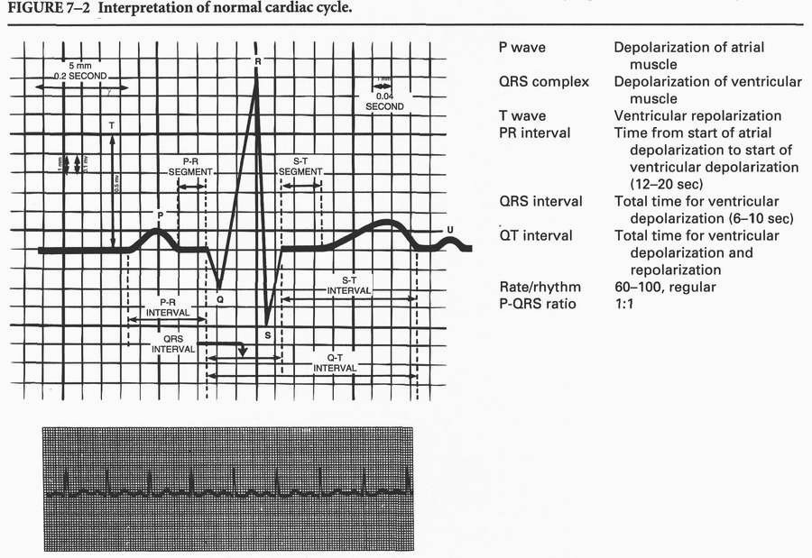
Comparison of Selected Cardiac Dysrhythmias
| Dysrhythmia | Description | Etiology | Symptoms/Consequences | Treatment |
| Dysrhythmias of Sinus Node | ||||
| Sinus dysrhythmia | Phasic shortening then lengthening of P-P and R-R interval | Respiratory variation in impulse initiation by SA node | Usually none | Usually none Atropine if rate below 40 beats per minute |
|
P waves present followed by QRS Rhythm regular Heart rate 100-150 beats per minute |
Increased metabolic demands Decreased oxygen delivery, heart failure, shock, hemorrhage, anemia |
May produce palpitations Prolonged episodes may lead to decreased cardiac output |
Treat underlying cause Occasionally sedatives |
|
| Sinus bradycardia |
P waves present followed by QRS Rhythm regular Heart rate <60 beats per minute |
Physical fitness Parasympathetic stimulation (sleep) Brain lesions |
Very low rates may cause decreased cardiac output light-headedness, faintness, chest pain |
Atropine if cardiac
output is decreased
Pacemaker Treat underlying cause |
Atrial Dysrhythmias |
||||
| Premature atrial |
QRS may or may not be normal Rhythm irregular |
Stress, ischemia, atrial enlargement, caffeine. |
May produce palpitations Frequent episodes may decrease cardiac output Is sign of chamber irrita- |
Sedation Quinidine May require no other treatment |
| Atrial tachycardia | P wave present (may merge into previous T wave), QRS usually normal; rapid heart rate usually >150 beats/minute | Sympathetic stimulation, chemical stimuli (caffeine nicotine), drug, toxicity |
Palpitations Possible anxiety |
Usually none Prolonged episodes may require carotid artery pressure, vagal stimulation, verapamil, digitalis, or beta blockers |
| Atrial fibrillation |
Rapid, irregular P waves (>350/minute) Ventricular rhythm irregularly irregular Ventricular rate varies, may increase to 120-150 if untreated |
Rheumatic heart disease |
Palpitations Pulse deficit Decreased cardiac output if rate is rapid Promotes thrombus formation in atria |
Digitalis Quinidine Cardioversion |
Ventricular Dysrhythmias |
||||
| Premature ventricular beats(PVBs) |
Earlv wide bizarre QRS, not associated with a P wave Rhythm irregular |
Stress, acidosis, ventricular enlargement Electrolyte imbalance Myocardial infarction Digitalis toxicity Hypoxemia, hypercapnia |
Same as for premature atrial beats |
MEDS:
|
| Ventricular tachycardia | No P wave before QRS; QRS wide and bizarre; ventricular rate > 100, usually 140-240 | PVB striking during vulnerable period; hypoxemia; drug toxicity; electrolyte imbalance; bradycardia | Decreased cardiac output, hypotension, loss of consciousness, respiratory arrest |
MEDS: Lidocaine Procainamide Bretylium Mexiletine Cardioversion Defibrillation |
| Ventricular fibrillation |
Chaotic electrical activity No recognizable QRS complex |
Myocardial infarction Electrocution Freshwater drowning Drug toxicity |
No cardiac output Absent pulse or respiration Cardiac arrest |
MEDS:
|
| Pulseless electrical activity (PEA) | Organized ECG rhythm |
Electromechanical dissociation |
Pulseless Minimal or no perfusion |
CPR Epinephrine |
| Ventricular standstill |
Can be distinguished from ventricular fibrillation only by ECG P waves may be present No QRS “Straight line” |
Myocardial infarction Chronic diseases of conducting system |
Same as for ventricular fibrillation |
CPR Pacemaker Intracardiac epinephrine Atropine |
Impulse Conduction |
||||
| First-degree atrioventricular (AV) block |
PR interval prolonged. |
Rheumatic fever Digitalis toxicity Degenerative changes of coronary atherosclerotic heart disease Infections (e.g., Lyme carditis) Decreased oxygen in AV node |
Warns of impaired conductions |
Usually none as long as it occurs as an isolated deficit Atropine if PR interval >0.26 second or bradycardia |
| Bundle branch block | Same as normal sinus rhythm (NSR) except QRS | Hypoxia, acute myocar-dial infarction, heart failure, coronary atherosclerotic heart disease, pulmonary embolus, hypertension | Same as first-degree AV | Usually none unless severe blockage of left posterior division |
| Second-degree AV blocks | P waves usually occur regularly at rates consistent with SA node initiation (not all P waves followed by QRS; PR interval may lengthen before nonconducted P wave or may be consistent; QRS may be widened) |
Acute myocardial infarction Same as first-degree AV block |
Serious dysrhythmia that may lead to decreased heart rate and cardiac output. |
May require temporary peacemaker If symptomatic (e.g., dizziness) atropine 1 mg. |
| Complete third-degree AV block |
Atria and ventricles beat independently P waves have no relation to QRS Ventricular rate may be as low as 20-40 beats per minute |
Digitalis toxicity Infectious disease Coronary artery disease Myocardial infarction |
Very low rates may cause decreased cardiac output: light-headedness, fainting, chest pain |
Pacemaker Isoproterenol to increase heart rate Epinephrine if isoproterenol ineffective |
- Cardiac arrest: sudden unexpected cessation of heartbeat and effective circulation leading to inadequate perfusion and sudden death.
- Risk factors:
- Myocardial infarction.
- Multiple traumas.
- Respiratory arrest.
- Drowning.
- Electrical shock.
- Drug reactions.
- Assessment - objective data:
- Unresponsive to stimuli (i.e., verbal, painful).
- Absence of breathing, carotid pulse.
- Pale or bluish: lips, fingernails, skin.
- Pupils: dilated.
- Analysis/nursing diagnosis:
- Decreased cardiac output related to heart failure.
- Impaired gas exchange related to breathlessness.
- Altered tissue perfusion related to pulselessness.
- Nursing care plan/implementation:
- Goal: prevent irreversible cerebral anoxic damage: initiate CPR within 4-6 minutes; continue until relieved; document assessment factors, effectiveness of actions; presence or absence of pulse at 1 minute and every 4 to 5 minutes.
- Goal: establish effective circulation, respiration.
- Evaluation/outcome criteria:
- Carotid pulse present; check after 1 minute and every few minutes thereafter.
- Responds to verbal stimuli.
- Pupils constrict in response to light.
- Return of spontaneous respiration; adequate ventilation.
- Arteriosclerosis: loss of elasticity, thickening, hardening of arterial walls; symptoms depend on organ system involved; common type - atherosclerosis. Atherosclerosis (coronary heart disease) precedes angina pectoris and myocardial infarction.
- Pathophysiology:
- Atherosclerotic plaque, discrete lumpy thickening of arterial wall; cholesterol-lipidcalcium deposits in lining.
- Narrows lumen, can occlude vessel.
- Risk factors:
- Increased serum cholesterol (low-density lipids 2:160 mg/dL).
- Hypertension.
- Cigarette smoking.
- Diabetes mellitus.
- Family history of premature CHD.
- Angina pectoris: transient paroxysmal episodes of substernal or precordial pain. Types: stable (follows an event, same severity); unstable (at rest or minimal exertion, recent onset, increasing severity); Prinzmetal’s variant (at rest, caused by coronary spasms).
- Pathophysiology:
- Insufficient blood flow through coronary arteries. Oxygen demand exceeds supply.
- Temporary myocardial ischemia.
- Risk factors:
- Cardiovascular:
- Atherosclerosis.
- Thromboangiitis obliterans.
- Aortic regurgitation.
- Hypertension.
- Hormonal:
- Hypothyroidism.
- Diabetes mellitus.
- Blood disorders:
- Anemia.
- Polycythemia vera.
- Lifestyle choices:
- Smoking.
- Obesity.
- Cocaine use.
- Inactivity.
- Assessment:
- Subjective data
- Pain.
- Type: squeezing, pressing, burning.
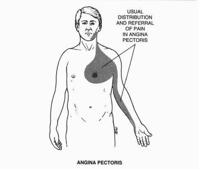
- Location: retrosternal, substernal, left of sternum, radiates to left arm.
- Duration: short, usually 3-5 minutes, <30 minutes
- Cause: emotional stress, overeating, physical exertion, exposure to cold; may occur at rest.
- Relief: rest, nitroglycerin.
- Dyspnea.
- Palpitations.
- Dizziness; faintness.
- Epigastric distress; indigestion; belching.
- Objective data
- Tachycardia.
- Pallor.
- Diaphoresis.
- ECG changes during attack.
- Analysis/nursing diagnosis:
- Altered cardiopulmonary tissue perfusion related to insufficient blood flow.
- Pain related to myocardial ischemia.
- Activity intolerance related to onset of pain.
- Nursing care plan/implementation:
- Goal: provide relief from pain.
- Rest until pain subsides.
- Nitroglycerin or amyl nitrite, beta-adrenergic blockers, as ordered.
- Identify precipitating factors: large meals, heavy exercise, stimulants (coffee, smoking), sex when fatigued, cold air.
- Vital signs: hypotension.
- Assist with ambulation; dizziness, flushing occurs with nitroglycerin.
- Goal: provide emotional support.
- Encourage verbalization of feelings, fears,
- Reassurance; positive self-concept.
- Acceptance of limitations.
- Goal: health teaching.
- Pain: alleviation, differentiation of angina from myocardial infarction, precipitating factors.
- Medication: frequency, expected effects (headache, flushing); carry fresh nitroglycerin; loses potency after 6 months (“stings” under tongue when potent); may use nitroglycerin paste-instruct how to apply.
- Diet: restricted calories if weight loss indicated; restricted fat, cholesterol, gasproducing foods; small, frequent meals.
- Diagnostic tests if ordered (e.g., thallium stress test, cardiac catheterization).
- Exercise: regular, graded, to promote coronary circulation.
- Prepare for coronary bypass surgery, if necessary.
- Behavior modification to assist with lifestyle changes, e.g., stress reduction, stop smoking.
- Evaluation/outcome criteria:
- Relief from pain.
- Fewer attacks.
- No myocardial infarction.
- Alters lifestyle; complies with limitations.
- No smoking.
Angina pectoris.
- Myocardial infarction (MI, heart attack): localized area of necrotic tissue in myocardium from cessation of blood flow; leading cause of death in North America.
- Pathophysiology:
- Coronary occlusion due to thrombosis, embolism, or hemorrhage adjacent to atherosclerotic plaque.
- Insufficient blood flow from cardiac hypertrophy, hemorrhage, shock, or severe dehydration.
- Risk factors:
- Age (35-70 year).
- Men more than women until menopause.
- Lifestyle: obesity, smoking, sedentary, amphetamine or cocaine use.
- Stress or type A personality.
- High-cholesterol, low-density lipoproteins, and serum triglyceride levels.
- Chronic illness (diabetes, hypertension).
- Assessment:
- Subjective data
- Pain
- Type: sudden, severe, crushing, heavy tightness. May be absent in elderly or diabetics.
- Location: substernal; radiates to one or both arms, jaw, neck. May be confused with indigestion.
- Duration: >30 minutes.
- Cause: unrelated to exercise; frequently occurs when sleeping (REM stage).
- Relief: oxygen, narcotics; not relieved by rest or nitroglycerin.
- Nausea.
- Shortness of breath.
- Apprehension, fear of impending death.
- History of cardiac disease (family); occupational stress.
- Objective data
- Vital signs: shock; rapid (>100), thready pulse; fall in blood pressure; S3 gallop; tachypnea, shallow respirations; elevated temperature within 24 hours (100°-103°F).
- Skin: ashen or clammy; diaphoretic.
- Emotional: restless.
- Lab data: increased - WBC (12000 - 15000/p,L), serum enzymes (CK-MB, LDH, > LDH2 - “flipped LDH”); changes - ECG (elevated ST segment, inverted T wave, arrhythmia).
Comparison of Physical Causes of Chest Pain
| Characteristic | Myocardial Infarction |
Pericarditis | Gastric Disorders | Angina Pectoris | Dissecting Aneurysm | Pulmonary Embolism |
| Onset | Gradual or sudden | Sudden | Gradual or sudden | Gradual or sudden | Abrupt, without prodromal symptoms | Gradual or sudden |
| Precipitating factors |
Can occur at rest or af- ter exercise or emotional stress |
Breathing deeply, rotating trunk, recumbency, swallowing or yawning |
Inflammation of stomach or esophagus; hypersecretion of gastric juices; some medications |
Usually after physical exertion, emotional stress,eating, exposure to cold,or defecation; unstable angina occurs at rest | Hypertension | Immobility or prolonged bedrest following surgery, trauma, hip fracture. HF, malignancy, oral contraceptives |
| Location | Substcrnal, anterior chest, or midline; rarely back; radiates to jaw or neck |
Precordial; radiates to neck or left shoulder and arm |
Xiphoid to umbilicus | Substernal, anterior chest; poorly localized |
Correlates with site of intimal rupture; anterior chest or back; between shoulder blades |
Pleural area, retrostenal area |
| Quality | Crushing, burning, stabbling, squeezing or vicelike |
Pleuritic, sharp | Aching, burning, cramp like, gnawing |
Squeezing, feeling of heavy pressure; burning |
Sharp, tearing or ripping sensation |
Sharp, stabbing |
| Intensity | Asymptomatic to severe; increases with time |
Mild to severe | Mild to severe | Mild to moderate | Severe and unbearable; maximal from onset | Aggravated by breathing |
| Duration | 30 minutes to 1-2 hours; may wax and wane |
Continuous | Periodic | Usually 2-10 minutes; average 3-5 minutes |
Continuous; docs not abate once started |
Variable |
| Relief | Narcotics | Sitting up, leaning forward | Physical and emotional rest, food, antacids, H2;receptor antagonists |
Nitroglycerin, rest | Large, repeated doses of narcotics | O2; sitting up morphine |
| Associated symptoms | Nausea, fatigue, heartbum; peripheral pulses equal |
Fever, dyspnea, nausea, anorexia, anxiety |
Nausea, vomiting, dysphagia, anorexia, weight loss | Belching, indigestion, dizziness |
Syncope, loss of sensations or pulses, oliguria; discrepancy between BP in arms; decrease in femoral or carotid pulse |
Dyspnea, tachypnea, diaphoresis, hemoptysis, cough, apprehension |
- Analysis/nursing diagnosis:
- Decreased cardiac output related to myocardial damage.
- Impaired gas exchange related to poor perfusion, shock.
- Pain related to myocardial ischemia.
- Activity intolerance related to pain or inadequate oxygenation.
- Fear related to possibility of death.
- Nursing care plan/implementation
- Goal: reduce pain, discomfort.
- Narcotics - morphine, possibly meperidine HCl (Demerol) or hydromorphone (Dilaudid); note response. Avoid IM.
- Humidified oxygen 2-4 L/min; mouth care - oxygen is drying.
- Position: semi-fowler’s to improve ventilation.
- Goal: maintain adequate circulation, stabilize heart rhythm.
- Monitor vital signs and urine output; observe for cardiogenic shock.
- Monitor ECG for arrhythmias.
- Give medications as ordered: antiarrhythmics - lidocaine HCl, atropine, quinidine HCl, procainamide (Pronestyl), bretylium (Bretylol); propranolol (Inderal); verapamil; anticoagulants - heparinsodium, bishydroxycoumarin or dicoumarin; thrombolytic agents - streptokinase (tPA), APSAC/anistreplase (Eminase).
- Recognize heart failure: edema, cyanosis, dyspnea, cough, crackles.
- Check lab data - normal; serum enzymes (CK 20-220 IU/L depending on gender; CK-MB 0-12 IU/L; LDH <115 IU/L; LDH1 < LDH2); blood gases (pH 7.35-7.45; Pco2 35-45 mEq/L; PO2 80-100 mm Hg; HCO3 22-26); electrolytes (K+ 3.5-5.0 mEq/L); clotting time (aPTT 25-41 seconds; PT 11-15 seconds).
- CVP - zero level at right atrium; fluctuates with respiration; normal range 5-15 cm H2O; note trend; increases with heart failure.
- ROM of lower extremities; TED hose/ antiembolic stockings.
- Goal: decrease oxygen demand/promote oxygenation, reduce cardiac workload.
- O2 as ordered.
- Activity: bed rest (24-48 hours) with bedside commode; planned rest periods; control visitors.
- Position: semi-Fowler’s to facilitate lung expansion and decrease venous return.
- Anticipate needs of patient: call light, water.
- Assist with feeding, turning.
- Environment: quiet, comfortable.
- Reassurance; stay with anxious patient.
- Give medications as ordered: cardiotonics, calcium channel blockers, vasodilators, vasopressors.
- Goal: maintain fluid electrolyte, nutritional status.
- IV (keep vein open); CVP; vital signs; urine output - 30 mL/hr.
- Lab data within normal limits (Na+ 135-145 mEq/L; K+ 3.5-5.0 mEq/L).
- Monitor ECG - hyperkalemia: peaked T wave; hypokalemia: depressed T wave.
- Diet: progressive low calorie, low sodium, low cholesterol, low fat, without caffeine.
- Goal: facilitate fecal elimination.
- •Da. Medications: stool softeners to prevent Valsalva maneuver (straining); mouth breathing during bowel movement; recognize complications of Valsalva maneuver - chest pain, cyanosis, diaphoresis, arrhythmias.
- Bedside commode if possible.
- Goal: provide emotional support.
- Recognize fear of dying: denial, anger, withdrawal.
- Encourage expression of feelings, fears, concerns.
- Discuss rehabilitation, lifestyle changes: prevent cardiac-invalid syndrome by promoting self-care activities, independence.
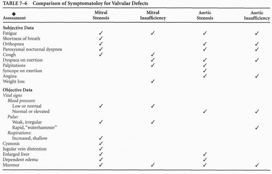
- Cardiac valvular defects: alteration in the structure of a valve; impede flow of blood or permit regurgitation.
- Pathophysiology:
- Stenosis - narrowing of valvular opening due to adherence, thickening, and rigidity of valve cusp from fibrosis, scarring, and calcification.
- Insufficiency (incompetence) - incomplete
- closure of valve due to contraction of chordae tendineae, papillary muscles; or to calcification, scarring of leaflets. Results in regurgitation.
- Mitral stenosis:
- Most common residual cardiac lesion of rheumatic fever.
- Affects women <45 yr more often than men.
- Narrowing of mitral valve.
- Interferes with filling of left ventricle.
- Produces pulmonary hypertension, right ventricular failure.
- Mitral insufficiency:
- Leaking/regurgitation of blood back into left atrium.
- Results from rheumatic fever, bacterial endocarditis; less common.
- Affects men more often.
- Produces pulmonary congestion, right ventricular failure.
- Aortic stenosis:
- Fusion of valve flaps between left ventricle and aorta.
- Congenital or acquired from atherosclerosis or from rheumatic fever and bacterial endocarditis; seen in men more often; pulmonary circulation congested, cardiac output decreased.
- Aortic insufficiency:
- Incomplete closure of valve between left ventricle and aorta (regurgitation).
- Left ventricular failure leading to right ventricular heart failure.
- Risk factors:
- Congenital abnormality.
- History of rheumatic fever.
- Atherosclerosis.
- Assessment:
- Analysis/nursing diagnosis:
- Decreased cardiac output related to inadequate ventricular filling.
- Fluid volume excess related to compensatory response to decreased cardiac output.
- Impaired gas exchange related to pulmonary congestion.
- Activity intolerance related to impaired cardiac function.
- Fatigue related to poor oxygenation.
- Nursing care plan/implementation:
- Goal: reduce cardiac workload.
- Goal: promote physical comfort and psychological support.
- Goal: prevent complications.
- Goal: prepare for surgery (commissurotomy, valvuloplasty [valvotomy], or valvular replacement, depending on defect and severity of condition).
- See section on cardiac surgery.
- Relief of symptoms.
- Increase in activity level.
- No complications following surgery.
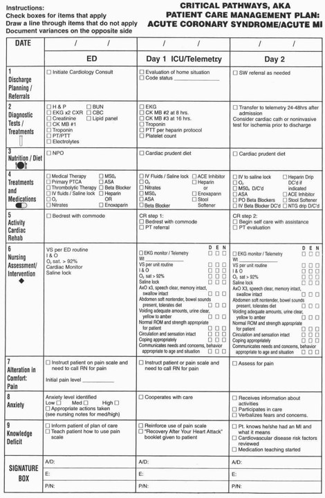
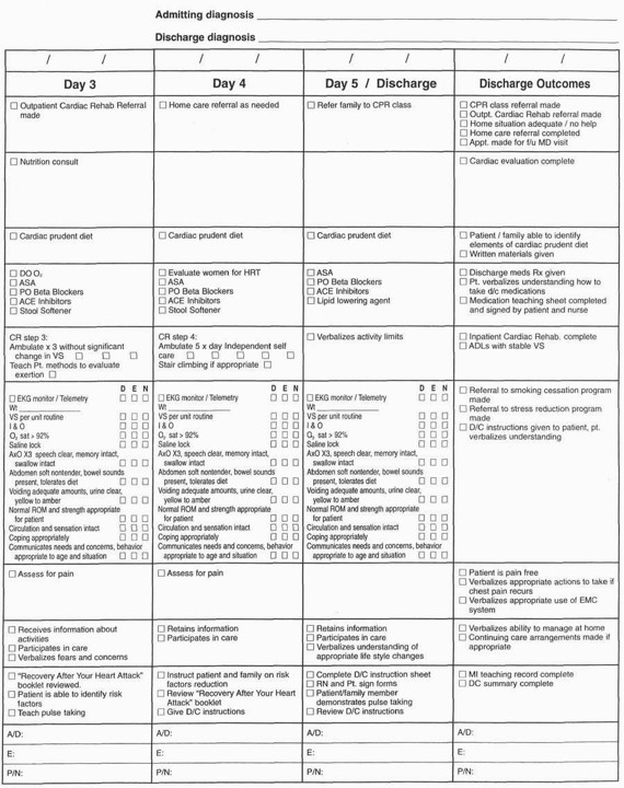
- Cardiac catheterization: a diagnostic procedure to evaluate cardiac status. Introduces a catheter into the heart, blood vessels; analyzes blood samples for oxygen content, cardiac output, pulmonary artery blood flow; done before heart surgery; frequently combined with angiography to visualize coronary arteries; also provides access for specialized cardiac techniques (e.g., internal pacing and coronary angioplasty).
- Approaches
- Right-heart catheterization - venous approach (antecubital or femoral) —> right atrium —> right ventricle —> pulmonary artery.
- Left-heart catheterization - retrograde approach: right brachial artery or percutaneous puncture of femoral artery —> ascending aorta —»left ventricle.
- Transseptal: femoral vein —> right atrium —> septum —> left atrium —> left ventricle.
- 0 b. Angiography/arteriography: done during left-heart catheterization.
- Precatheterization
- Assessment:
- Subjective data
- Allergies: iodine, seafood.
- Anxiety.
- Objective data
- Vital signs: baseline data.
- Distal pulses: mark for reference after catheterization.
- Analysis/nursing diagnosis:
- Anxiety related to fear of unknown.
- Knowledge deficit (learning need) related to limited exposure to information or sudden need for procedure.
- Nursing care plan/implementation:
- Goal: provide for safety, comfort.
- Signed informed consent.
- NPO (except for medications 6-8 hours before).
- Have patient urinate before going to lab.
- Give sedatives, as ordered, 30 minutes before procedure (e.g., midazolam HCl [Versed] IV, diazepam [Valium] PO).
- Goal: health teaching.
- Procedure: length (1-3 hours).
- Expectations (strapped to table for safety, must he still, awake but mildly sedated).
- Sensations (hot, flushed feeling in head with dye injection; thudding in chest from premature beats during catheter manipulation; desire to cough, particularly with right-heart angiography and contrast-medium injection).
- Alert physician to unusual sensations (coolness, numbness, paresthesia).
- Postcatheterization
- Assessment (potential complications):
- Subjective data
- Puncture site: increasing pain, tenderness.
- Palpitations.
- Affected extremity: tingling, numbness, pain from hematoma or nerve damage.
- Objective data
- Vital signs: shock, respiratory distress (related to pulmonary emboli, allergic reaction).
- Puncture site: bleeding (hematoma).
- ECG: arrhythmias, signs of MI.
- Affected extremity: color, temperature, peripheral pulses.
- Analysis/nursing diagnosis:
- Decreased cardiac output related to arrhythmias or MI.
- Altered tissue perfusion related to bleeding following procedure.
- Pain related to puncture site tenderness.
- Nursing care plan/implementation:
- Goal: prevent complications.
- Bed rest: depends on size of catheter and closure procedure - Perclose dissolvable suture, 30 minute; Angioseal (collagen plug), 2 hours; compression pump or 15 minutes manual compression followed by sandbag, 4-5 hours; 12-24 hours with sheath or antiplatelet drip (abciximab ReoPro).
- Vital signs: record ql5min for 1 hour, q30 minutes for 3 hours or until stable; check BP on opposite extremity.
- Puncture site: observe for bleeding, swelling, or tenderness; check pulse distal to insertion site to determine patency of artery; report complaints of coolness, numbness, or paresthesia in extremity.
- ECG: monitor, document rhythm.
- Give medications as ordered: sedatives; mild narcotics; antiarrhythmics; antiplatelet (Plavix, aspirin) or lowmolecular-weight heparin (enoxaparin [Lovenox]) with stent insertion.
- Goal: provide emotional support.
- Explanations: brief, accurate; patient anxious to learn results of test.
- Counseling: refer as indicated.
- Goal: health teaching.
- Late complications: infection.
- Prepare for surgery if indicated.
- Follow-up medical care.
- Limitations following PTCA: no lifting >10 lb and no vigorous exertion for 1-2 weeks; return to normal work and sexual activity in 2-3 days.
- Evaluation/outcome criteria: no complications (e.g., cardiac arrest, hematoma at insertion site).
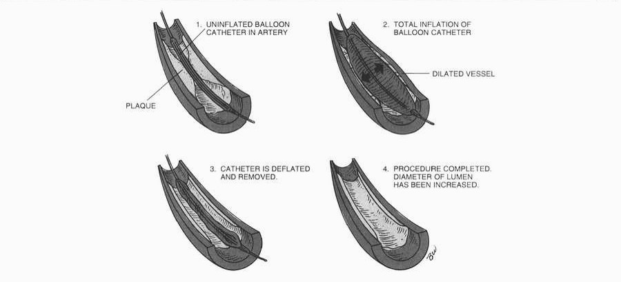
- Percutaneous transluminal coronary angioplasty (PTCA): a balloon-tipped catheter is threaded to site of coronary occlusion and inflated repeatedly until blood flow increases distal to the obstruction; a nonsurgical alternative to bypass surgery for coronary artery occlusion; recommended in patients with poorly controlled angina, mild or no symptoms, multiple- or single-vessel disease with a noncalcified, discrete, and proximal lesion that can be reached by the catheter; costs less and requires shorter hospitalization and rehabilitation period; successful in 90% of patients; approximately 30% restenose by 3 months. Rotational atherectomy may also be done; a high-speed drill pulverizes plaque into small particles. An intravascular stent, steel mesh or coil spring, may be placed in the coronary artery; the stent acts as a mechanical scaffold to reopen the blocked artery. The patient receives low-molecular-weight heparin and/or platelet therapy following the procedure.
Arterial balloon angioplasty.
 |
-
Cardiac surgery: done to alter the structure of the heart or vessels when congenital or acquired disorders interfere with cardiac functioning: septal defects; transposition of great vessels; tetralogy of Fallot; pulmonary/aortic stenosis; coronary artery bypass; valve replacement.
Cardiopulmonary bypass (open-heart surgery): blood from cardiac chambers and great vessels is diverted into a pump oxygenator; allows full visualization of heart during surgery; maintains perfusion and body functioning. - Preoperative
- Assessment: see specific conditions for preoperative signs and symptoms, i.e., valvular defects, angina, MI; Establish complete baseline: daily weight; vital signs - integrity of all pulses, BP both arms; CVP or pulmonary artery pressures (Swan-Ganz); neurological status; emotional status; nutritional and elimination patterns; laboratory values (urine, electrolytes, enzymes, coagulation studies); pulmonary function studies.
- Analysis/nursing diagnosis:
- Decreased cardiac output related to myocardial damage.
- Activity intolerance related to poor cardiac function.
- Knowledge deficit (learning need) related to insufficient time for teaching.
- Anxiety related to fear of unknown.
- Fear related to possible death.
- Risk for spiritual distress related to possible death.
- Nursing care plan/implementation:
- Goal: provide emotional and spiritual support.
- Arrange for religious consultation if desired.
- Provide opportunity for family visit morning of surgery.
- Encourage verbalization/questions: fear, depression, despair frequently occur.
- Goal: health teaching.
- Diagnostic procedures, treatments, specifics for surgery (i.e., internal mammarian artery or leg incision with use of saphenous vein in coronary artery bypass surgery).
- Postoperative regimen: turn, cough, deep breathe, ROM, equipment used, medication for pain.
- Tour ICU; meet personnel.
- Alternative method of communication while intubated.
- Evaluation/outcome criteria:
- Displays moderate anxiety level.
- Verbalizes/demonstrates postoperative expectations.
- Quits smoking before surgery.
- Postoperative
- Assessment:
- Subjective data
- Pain.
- Fatigue - sleep deprivation.
- Objective data
- Neurological: level of consciousness; pupillary reactions; movement of limbs (purposeful, spontaneous).
- Respiratory: rate changes (increases occur with obstruction, pain; decreases occur with CO2 retention); depth (shallow with pain, atelectasis); symmetry; skin color; patency/drainage from chest tubes, sputum (amount, color); endotracheal tube placement (bilateral breath sounds).
- Cardiovascular:
- BP - hypotension may indicate heart failure, tamponade, hemorrhage, arrhythmias, or thrombosis; hypertension may indicate anxiety, hypervolemia.
- Pulse: radial, apical, pedal; rate (>100 may indicate shock, fever, hypoxia, arrhythmias); rhythm, quality.
- CVP or Swan-Ganz (elevated in cardiac failure); temperature (normal postop: 98.6°-101.6°F oral).
- GI: nausea, vomiting, distention.
- Renal: urine - minimum output (30 mL/hour); color;specific gravity (<1.010 occurs with overhydration, renal tubular damage; >1.020 present with dehydration, oliguria, blood in urine).
- Analysis/nursing diagnosis:
- Decreased cardiac output related to decreased myocardial contractility or postoperative hypothermia.
- Pain related to incision.
- Inejfective airway clearance related to effects of general anesthesia.
- Altered tissue perfusion related to postoperative bleeding or thromboemboli.
- Fluid volume deficit related to blood loss.
- Risk for infection related to wound contamination.
- Altered thought processes related to anesthesia or stress.
- Altered role performance related to uncertainty about future.
- Nursing care plan/implementation:
- Goal: provide constant monitoring to prevent complications.
- Respiratory:
- Observe for respiratory distress: restlessness, nasal flaring, CheyneStokes respiration, dusky/cyanotic skin; assisted or controlled ventilation via endotracheal tube common 6-24 hours; supplemental O2 after extubation.
- Suctioning; cough, deep breathe.
- Elevate head of bed.
- Position chest tube to facilitate drainage; mediastinal sump tube maintains patency - “milking” not necessary.
- Cardiovascular:
- Vital signs: BP >80-90 mm Hg systolic; CVP: range 5-15 cm H2O unless otherwise ordered; pulmonary artery line (Swan-Ganz): mean pressure 4-12 mm Hg; I&O: report <30 mL/hr of urine from indwelling urinary catheter.
- ECG; PVCs occur most frequently following aortic valve replacement and bypass surgery.
- Peripheral pulses if leg veins used for grafting.
- Activity: turn q2h; ROM; progressive, early ambulation.
- Inspect dressing for bleeding.
- Medications according to therapeutic directives - cardiotonics (digoxin);coronary vasodilators (nitrates); antibiotics (penicillin); analgesics; anticoagulants (with valve replacements); antiarrhythmics (quinidine, procainamide HC1 [Pronestyl]).
- Goal: promote comfort, pain relief.
- Medicate: Demerol or morphine sulfate - severe pain lasts 2-3 days.
- Splint incision when moving or coughing.
- Mouth care: keep lips moist.
- Position: use pillows to prevent tension on chest tubes, incision.
- Goal: maintain fluid, electrolyte, nutritional balance.
- I&O; urine specific gravity.
- Measure chest drainage - should not exceed 200 mL/hour for first 4-6 hours.
- Give fluids as ordered; maintain IV patency.
- Diet: clear fluids —» solid food if no nausea, GI distention; sodium intake restricted, low fat.
- Goal: promote emotional adjustment.
- Anticipate behavior disturbances (depression, disorientation often occur 3 days postop) related to medications, fear, sleep deprivation.
- Calm, oriented, supportive environment, as personalized as possible.
- Encourage verbalization of feelings (family and patient).
- Encourage independence to avoid cardiac-cripple role.
- Goal: health teaching.
- Alterations in lifestyle; activity, diet, work; resumption of sexual activity usually when patient can climb two flights of stairs.
- Available community resources for cardiac rehabilitation (e.g., American Heart Association, Mended Hearts).
- Drug regimen: purpose, side effects.
- Potential complications: dyspnea, pain, palpitations common postoperatively.
- Evaluation/outcome criteria:
- No complications; incision heals.
- Activity level increases - no signs of overexertion (e.g., fatigue, dyspnea, pain).
- Relief of symptoms.
- Returns for follow-up medical care.
- Takes prescribed medications; knows purposes and side effects.
- Minimally invasive direct coronary artery bypass (MIDCAB): a variation of CABG for patients in whom sternotomy and cardiopulmonary bypass is contraindicated or unnecessary. The left internal mammary is anastomosed to the left anterior descending coronary artery through a thoracic incision without bypass.
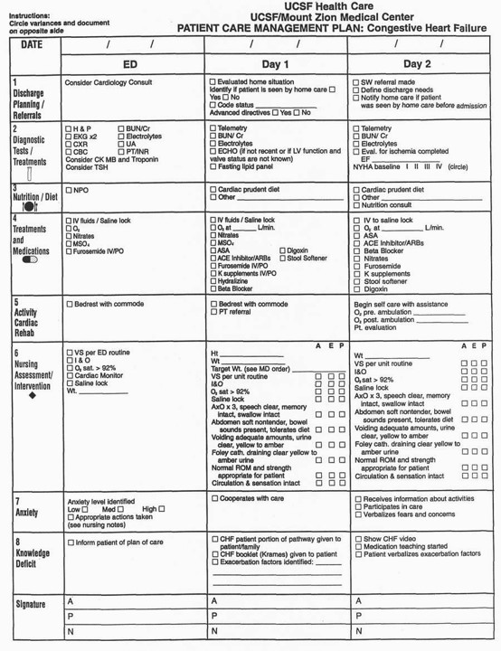
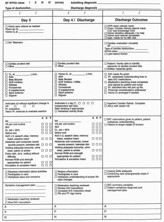
- Heart failure (HF): inability of the heart to meet the peripheral circulatory demands of the body; cardiac decompensation; combined right and left ventricular heart failure.
- Pathophysiology: increased cardiac workload or decreased effective myocardial contractility—» decreased cardiac output (forward effects). Left ventricular failure —» pulmonary congestion; right atrial and right ventricular failure —» systemic congestion —» peripheral edema (backward effects). Compensatory mechanisms in HF include tachycardia, ventricular dilation, and hypertrophy of the myocardium; develops in 50-60% of heart disease patients.
- Risk factors:
- Decreased myocardial contractility:
- Myocarditis.
- MI.
- Tachyarrhythmias.
- Bacterial endocarditis.
- Acute rheumatic fever.
- Increased cardiac workload:
- Elevated temperature.
- Physical/emotional stress.
- Anemia.
- Hyperthyroidism (thyrotoxicosis).
- Valvular defects.
- Assessment:
- Subjective data
- Shortness of breath.
- Orthopnea (sleeps on two or more pillows).
- Paroxysmal nocturnal dyspnea (sudden breathlessness during sleep).
- Dyspnea on exertion (climbing stairs),
- Apprehension; anxiety; irritability.
- Fatigue; weakness.
- Reported weight gain; feeling of puffiness.
- Objective data
- Vital signs:
- BP: decreasing systolic; narrowing pulse pressure.
- Pulse: pulsus alternans (alternating strong-weak-strong cardiac contraction), increased.
- Respirations: crackles, Cheyne-Stokes.
- Edema: dependent, pitting (1+ to 4+ mm).
- Liver: enlarged, tender
- Neck veins: distended.
- Chest x-ray:
- Cardiac enlargement.
- Dilated pulmonary vessels.
- Diffuse interstitial lung edema.
- Analysis/nursing diagnosis:
- Decreased cardiac output related to decreased myocardial contractility.
- Activity intolerance related to generalized weakness and inadequate oxygenation.
- Fatigue related to edema and poor oxygenation.
- Altered tissue perfusion related to peripheral edema and inadequate blood flow.
- Fluid volume excess related to compensatory mechanisms.
- Impaired gas exchange related to pulmonary congestion.
- Anxiety related to shortness of breath.
- Sleep pattern disturbance related to paroxysmal nocturnal dyspnea.
- Nursing care plan/implementation:
- Goal: provide physical rest/reduce emotional stimuli.
- Position: sitting or semi-fowler's until tachycardia, dyspnea, edema resolved; change position frequently; pillows for support.
- Rest: planned periods; limit visitors, activity, noise. Chair and commode privileges.
- Support: stay with anxious patient; have supportive family member present; administer sedatives/tranquilizers as ordered.
- Warm fluids if appropriate.
- Goal: provide for relief of respiratory distress; reduce cardiac workload.
- Oxygen: low flow rate; encourage deep breathing (5-10 minutes q2h); auscultate breath sounds for congestion, pulmonary edema.
- Position: elevating head of bed 20-25 cm (8-10 in) alleviates pulmonary congestion.
- Medications as ordered:
- Digitalis preparations.
- ACE inhibitors-captopril, enalapril.
- Inotropic agent-dobutamine, dopamine.
- Diuretics-thiazides, furosemide, metolazone.
- Tranquilizers-phenobarbital, diazepam (Valium), chlordiazepoxide (Librium) HC1.
- Vasodilators-hydrolazine, isosorbide.
- Goal: provide for special safety needs.
- Skin care:
- Inspect, massage, lubricate bony prominences.
- Use foot cradle, heel protectors; sheepskin.
- Side rails up if hypoxic (disoriented).
- Vital signs: monitor for signs of fatigue, pulmonary emboli.
- ROM: active, passive; elastic stockings.
- Goal: maintain fluid and electrolyte balance, nutritional status.
- Urine output: 30 mL/hr minimum; estimate insensible loss in diaphoretic patient. Monitor BUN, serum creatinine, and electrolytes.
- Daily weight; same time, clothes, scale.
- IV: use microdrip to avoid circulatory overloading.
- Diet:
- Low sodium as ordered.
- Small, frequent feedings.
- Discuss food preferences with patient.
- Goal: health teaching.
- Diet restrictions; meal preparation.
- Activity restrictions, if any; planned rest periods.
- Medications: schedule (e.g., diuretic in early morning to limit interruption of sleep) purpose, dosage, side effects (importance of daily pulse taking, daily weights, intake of potassium-containing foods).
- Available community resources for dietary assistance, weight reduction, exercise program.
- Evaluation/outcome criteria:
- Increase in activity level tolerance - fatigue decreased.
- No complications - pulmonary edema, respiratory distress.
- Reduction in dependent edema.
Left Ventricular Compared with Right Ventricular Heart Failure
|
Left Ventricular Failure |
Right Ventricular Failure |
|
|
- Pulmonary edema: sudden transudation of fluid from pulmonary capillaries into alveoli. Lifethreatening condition.
- Pathophysiology: increased pulmonary capillary permeability; increased hydrostatic pressure (pulmonary hypertension); decreased blood colloidal osmotic pressure; fluid accumulation in alveoli —> decreased compliance —> decreased diffusion of gas —> hypoxia, hypercapnea.
- Risk factors:
- Left ventricular failure.
- Pulmonary embolism.
- Drug overdose.
- Smoke inhalation.
- CNS damage.
- Fluid overload.
- Assessment:
- Subjective data
- Anxiety.
- Restlessness at onset progressing to agitation.
- Stark fear.
- Intense dyspnea, orthopnea, fatigue.
- Objective data
- Vital signs:
- Pulse: tachycardia; gallop rhythm.
- Respiration: tachypnea, moist, bubbling, wheezing, labored.
- Temperature: normal to subnormal.
- Skin: pale, cool, diaphoretic, cyanotic
- Auscultation: crackles, wheezes.
- Cough: productive of large quantities of pink, frothy sputum.
- Right ventricular heart failure: distended (bulging) neck veins, peripheral edema, hepatomegaly, ascites.
- Mental status: restless, confused, stuporous.
- Arterial blood gases: hypoxia; pulse oximetry: decreased O2 saturation.
- Chest x-ray: haziness of lung fields, cardiomegaly.
- Analysis/nursing diagnosis:
- Decreased cardiac output related to decreased myocardial contractility.
- Impaired gas exchange related to pulmonary congestion.
- Altered tissue perfusion related to inadequate blood flow.
- Anxiety, severe, related to difficulty breathing.
- Fear related to life-threatening situation.
- Nursing care plan/implementation:
- Goal: promote physical, psychological relaxation measures to relieve anxiety.
- Slow respirations: morphine sulfate 3-10 mg IV/SC/IM, as ordered, to reduce respiratory rate, sedate, and produce vasodilation.
- Remain with patient.
- Encourage slow, deep breathing; assist with coughing.
- Work calmly, confidently, unhurriedly.
- Frequent rest periods.
- Goal: improve cardiac function, reduce venous return, relieve hypoxia.
- O2: 100%, preferably by demand valve to slow respiratory rate, provide uniform ventilation, reduce venous return, and inhibit “leaky capillary” syndrome. Possibly PEEP.
- IV:D5W
- Give aminophylline, as ordered, to lower venous pressure and increase cardiac output.
- Position: high Fowler’s, extremities in dependent position, to reduce venous return and facilitate breathing.
- Medications as ordered: digitalis; diuretics - furosemide (Lasix); inotropic agents -dobutamine (Dobutrex), dopamine; nitroglycerin, nitroprusside.
- Vital signs; auscultate breath sounds.
- Diet: low sodium; fluid restriction as ordered.
- Goal: health teaching (include family or significant other).
- Medications.
- Side effects.
- Potassium supplements if indicated.
- Pulse taking.
- Exercise; rest.
- Diet: low sodium.
- Signs of complications: edema; weight gain of 2-3 lb (0.9-1.4 kg) in a few days; dyspnea.
- Evaluation/outcome criteria:
- No complications; vital signs stable; clear breath sounds.
- No weight gain; weight loss if indicated.
- Alert, oriented, calm.
- Shock-: a critically severe deficiency in nutrients, oxygen, and electrolytes delivered to body tissues, plus deficiency in removal of cellular wastes; results from cardiac failure, insufficient blood volume, or increased vascular bed size.
- Types, pathophysiology, and risk factors:
- Hypovolemic (hemorrhagic, hematogenic) - markedly decreased volume of blood (hemorrhage or plasma loss from intestinal obstruction, burns, physical trauma, or dehydration) —> decreased venous return, cardiac output —> decreased tissue perfusion.
- Cardiogenic - failure of cardiac muscle pump (myocardial infarction) —> generally de-
- creased cardiac output —> pulmonary congestion, hypoxia —> inadequate circulation; high mortality rate.
- Distributive:
- Neurogenic - massive vasodilation from reduced vasomotor, vasoconstrictor tone (e.g., spinal shock, head injuries, anesthesia, pain); interruption of sympathetic nervous system; blood volume is normal but inadequate for vessels —> decreased venous return —> tissue hypoxia.
- Vasogenic (anaphylactic, septic, endotoxic)—severe reaction to foreign protein (insect bites, drugs, toxic substances, aerobic, gram-negative organisms) —> histamine release —> vasodilation, venous stasis —> diminished venous return.
- Assessment: varies, depending on degree of shock.
- Subjective data
- Anxiety; restlessness.
- Dizziness; fainting.
- Thirst.
- Nausea.
- Objective data
- Vital signs:
- BP - hypotension (postural changes in early shock; systolic <70 mm Hg in late shock).
- Pulse - tachycardia, thready; irregular (cardiogenic shock); could be slow if conduction system of heart damaged.
- Respirations - increased depth, rate; wheezing (anaphylactic shock).
- Temperature - decreased (elevated in septic shock).
- Skin:
- Pale (or mottled), cool, clammy (warm to touch in septic shock).
- Urticaria (anaphylactic shock).
- Level of consciousness: alert, oriented —> unresponsive.
- CVP
- Below 5 cm H2O with hypovolemic shock.
- Above 15 cm H2O with cardiogenic, possibly septic shock.
- Urine output: decreased (<30 mL/hr).
- Capillary refill: slowed; normally nailbed “pinks up” within 2 seconds after blanching (nailbed pressure).
- Analysis/nursing diagnosis:
- Altered tissue perfusion related to vasoconstriction or decreased myocardial contractility.
- Impaired gas exchange related to ventilationperfusion imbalance.
- Decreased cardiac output related to loss of circulating blood volume or diminished cardiac contractility.
- Altered urinary elimination related to decreased renal perfusion.
- Fluid volume deficit related to blood loss.
- Anxiety related to severity of condition.
- Risk for injury related to death.
- Nursing care plan/implementation: Goal: promote venous return, circulatory perfusion.
- Position: foot of bed elevated 20 degrees (12-16 inches), knees straight, trunk horizontal, head slightly elevated; avoid Trendelenburg position.
- Ventilation: monitor respiratory effort, loosen restrictive clothing; O2 as ordered.
- Fluids: maintain IV infusions; give blood, plasma expanders as ordered (exception - stop blood immediately in anaphylactic shock).
- Vital signs:
- CVP (decreased with hypovolemia) arterial line, Swan-Ganz (increased pulmonary artery wedge pressue indicating cardiac failure).
- Urine output (insert catheter for hourly output).
- Monitor ECG (increased rate, dysrhythmias).
- Medications (depending on type of shock) as ordered:
- Antihypotensives - epinephrine (Adrenalin), norepinephrine (Levophed), isoproterenol (Isuprel), dopamine (Intropin).
- Antiarrhythmics.
- Cardiac glycosides.
- Adrenocorticoids.
- Antibiotics.
- Vasodilators.
- Beta-adrenergies.
- Mechanical support: military (or medical) antishock trousers (MAST) or pneumatic antishock garment (PASG); used to promote internal autotransfusion of blood from legs and abdomen to central circulation; at lower pressures may control bleeding and promote hemostasis; do not remove (deflate) suddenly to examine underlying areas or BP will drop precipitously; compartment syndrome may result with prolonged use and high pressure; controversial.
- Evaluation/outcome criteria:
- Vital signs stable, within normal limits.
- Alert, oriented.
- Urine output >30 mL/hr.
Signs of Hypovolemic Shock
|
Blood Loss (% of Total) |
Assessment Data |
|
800-1500 mL (15-30%) |
|
|
2000 mL (30-40%) |
|
| >2500 mL (>40%) |
|
- Disseminated intravascular coagulation (DIC): diffuse or widespread coagulation initially within arterioles and capillaries leading to hemorrhage.
- Pathophysiology: activation of coagulation system from tissue injury —> fibrin microthrombi form in brain, kidneys, lungs —> microinfarcts, tissue necrosis —» red blood cells, platelets, prothrombin, other clotting factors trapped, destroyed in process —> excessive clotting —» release of fibrin split products —» inhibition of platelet clotting —» profuse bleeding.
- Risk factors:
- Obstetric complications.
- Neoplastic disease.
- Low perfusion states (e.g., burns, hypothermia).
- Infections.
- Assessment - objective data:
- Skin, mucous membranes: petechiae, ecchymosis.
- Extremities (fingers, toes): cyanosis.
- Bleeding: venipuncture sites, wound, oral, rectal, vaginal.
- Urine output: oliguria —> anuria.
- Level of consciousness: convulsions, coma.
- Lab data: prolonged - prothrombin time (PT) >15 seconds; decreased - platelets, fibrinogen level.
- Analysis/nursing diagnosis:
- Altered tissue perfusion related to peripheral microthrombi.
- Risk for injury related to bleeding.
- Risk for impaired skin integrity related to ischemia.
- Altered urinary elimination related to renal tubular necrosis.
- Nursing care plan/implementation: goal - prevent and detect further bleeding.
- Carry out nursing measures designed to alleviate underlying problem (e.g., shock, birth of fetus, surgery/irradiation for cancer, antibiotics for infection).
- Medications: heparin sodium IV, 1000 U/hr, if ordered, to reverse abnormal clotting (controversial).
- IVs: blood to lessen shock; platelets, cryoprecipitate, fresh plasma to restore clotting factors, fibrinogen.
- Observe: vital signs, CVP (normal 5-15 mm Hg), PAP (normal 20-30 systolic and 8-12 diastolic), and intake and output for signs of shock or fluid overload from frequent infusions; specimens for occult blood (urine, stool).
- Precautions: avoid IM injections if possible; pressure 5 minutes to venipuncture sites; no rectal temperatures.
- Evaluation/outcome criteria:
- Clotting mechanism restored (increased platelets, normal PT).
- Renal function restored (urine output >30 mL/hr).
- Circulation to fingers, toes; no cyanosis.
- No irreversible damage from renal, cerebral, cardiac, or adrenal hemorrhage.
- Pericarditis: inflammation of parietal or visceral pericardium or both; acute or chronic condition; may occur with or without effusion. Cardiac tamponade may result.
- Pathophysiology: fibrosis or accumulation of fluid in pericardium —» compression of cardiac pumping —»decreased cardiac output —> increased systemic, pulmonic venous pressure.
- Risk factors:
- Bacterial, viral, fungal infections.
- Tuberculosis.
- Collagen diseases.
- Uremia.
- Transmural MI.
- Trauma.
- Assessment:
- Subjective data
- Pain:
- Type - sharp, moderate to severe.
- Location - wide area of pericardium, may radiate: right arm, jaw/teeth.
- Precipitating factors - movement, deep inspiration, swallowing.
- Chills; sweating.
- Apprehension; anxiety.
- Fatigue.
- Abdominal pain.
- Shortness of breath.
- Objective data
- Vital signs:
- BP: decreased pulse pressure; pulsus paradoxus - abnormal drop in systolic BP of >8-10 mm Hg during inspiration.
- Pulse: tachycardia.
- Temperature: elevated; erratic course; low grade.
- Pericardial friction rub.
- Increased CVP; distended neck veins; dependent pitting edema; liver engorgement.
- Restlessness.
- jj e. Lab data: elevated AST (SGOT), WBC;
- CT or MR1 - pericardial thickening.
- 3 f. Serial ECGs: increased ST segment; echocardiogram: pericardial fluid.
- Analysis/nursing diagnosis:
- Decreased cardiac output related to impaired cardiac muscle contraction.
- Pain related to pericardial inflammation.
- Anxiety related to unknown outcome.
- Fatigue related to inadequate oxygenation.
- Nursing care plan/implementation:
- Goal: promote physical and emotional comfort.
- Position: semi-fowler’s (upright or sitting); bed rest.
- Vital signs: q2-4h and pm; apical and radial pulse; notify physician if heart sounds decrease in amplitude or if pulse pressure narrows, indicating cardiac tamponade; cooling measures as indicated.
- O2 as ordered.
- Medications as ordered:
- Analgesics—aspirin, morphine sulfate, meperidine or codeine.
- Nonsteroidal anti-inflammatory agents —indomethacin.
- Antimicrobial.
- Digitalis and diuretics, if heart failure present.
- Assist with aspiration of pericardial sac (pericardiocentesis) if needed: medicateB as ordered; elevate head 60 degrees; monitor ECG; have defibrillator and pacemaker available.
- Prepare for pericardiectomy (excision of constricting pericardium) as ordered.
- Continual emotional support.
- Enhance effects of analgesics: positioning; turning; warm drinks.
- Monitor for signs of cardiac tamponade: tachycardia; tachypnea; hypotension; pallor; narrowed pulse pressure; pulsus paradoxus; distended neck veins.
- Goal: maintain fluid, electrolyte balance.
- Parenteral fluids as ordered; strict I&O.
- Assist with feedings; low-sodium diet may be ordered.
- Evaluation/outcome criteria:
- Relief of pain, dyspnea.
- No complications (e.g., cardiac tamponade).
- Return of normal cardiac functioning.
- Chronic arterial occlusive disease: arteriosclerosis obliterans most common occlusive disorder of the arterial system (aorta, large and medium-size arteries); frequently involves the femoral, iliac, and popliteal arteries.
- Pathophysiology: fatty deposits in intimal, medial layer of arterial walls; plaque formation —> narrowed arterial lumens; decreased distensibility —> decreased blood flow; ischemic changes in tissues.
- Risk factors:
- Age (>50).
- Sex (men).
- Diabetes mellitus.
- Hyperlipidemia - obesity.
- Cigarette smoking.
- Hypertension.
- Family history.
- Assessment:
- Subjective data
- Pain:
- Type - cramplike.
- Location - foot, calf, thigh, buttocks.
- Duration - variable, may be relieved by rest.
- Precipitating causes - exercise (intermittent claudication), but occasionally may occur when at rest.
- Tingling, numbness in toes, feet.
- Persistent coldness of one or both lower extremities.
- Objective data
- Lower extremities:
- Pedal pulses - absent or diminished.
- Skin - shiny, glossy; dry, cold, chalky white, decreased/absent hair, ulcers, gangrene.
- Lab data: increased serum cholesterol, I triglycerides, CBC, platelets
- J c. Arteriography - indicates location, nature of occlusion. Noninvasive: ultrasound, segmental limb pressure, exercise testing.
- Analysis/nursing diagnosis:
- Altered tissue perfusion related to peripheral vascular disease.
- Risk for activity intolerance related to pain and sensory changes.
- Pain related to ischemia.
- Risk for impaired skin integrity related to poor circulation.
- Risk for injury related to numbness of extremities.
- Nursing care plan/implementation:
- Goal: promote circulation; decrease discomfort.
- Position: elevate head of bed on blocks (3-6 inches), because gravity aids perfusion to thighs, legs; elevating legs increases pain.
- Comfort: keep warm: avoid chilling or use of heating pads, which may burn skin; apply bed socks.
- Circulation: check pedal pulses, skin color, temperature qid.
- Medications:
- Vasodilators.
- Antiplatelet - ASA, tidopidine, dipyridamole.
- Dihydropyridines - nifedipine, amlodipine.
- Xanthine derivatives - pentoxifylline.
- Goal: prevent infection, injury.
- Skin care: use bed cradle, sheepskin, heel pads; mild soap; dry thoroughly; lotion; do not massage, to prevent release of thrombus.
- Foot care: wear properly fitting shoes, slippers when out of bed; inspect for injury or pressure areas; nail care by podiatrist.
- Goal: health teaching.
- Skin care; inspect daily.
- Activity: balance exercise, rest to increase collateral circulation; walk only until painful.
- Exercises: walking, Buerger-Allen exercises (gravity alternately fills and empties blood vessels).
- Diet: /ow-fat, heart-healthy diet to slow disease progression.
- Lifestyle choices: avoid smoking.
- Recognizes and reports signs of occlusion (e.g., pain, cramping, numbness in extremities, color changes - white or blue, temperature changes - cool to cold).
- Evaluation/outcome criteria:
- Decreased pain.
- Skin integrity preserved; no loss of limb.
- Quits smoking.
- Does exercises to increase collateral circulation.
- Aneurysms (thoracic or abdominal aortic): localized or diffuse dilations/outpouching of a vessel wall, usually an artery; exerts pressure on adjacent structures; affects primarily men over age 60; >6 cm diameter, 50% will rupture; resected surgically, reconstructed with synthetic or vascular graft.
- Risk factors:
- Atherosclerosis.
- Trauma.
- Syphilis.
- Congenital weakness.
- Local infection.
- Cigarette smoking.
- Assessment:
- Subjective data
- Pain:
- Constant, boring, neuralgic, intermittent - low back, abdominal.
- Angina - sudden onset may mean rupture or dissection, which are emergency conditions.
- Dyspnea; orthopnea - pressure on trachea or bronchus.
- Objective data
- Vital signs:
- Radial pulses differ.
- Tachycardia.
- Hypotension following rupture leading to shock.
- Pulsating mass: abdominal, chest wall pulsation; edema of chest wall (thoracic aneurysm); periumbilical (abdominal aneurysm); audible bruit over aorta.
- Cyanosis, mottled below level of aneurysm.
- Veins: dilated, superficial - neck, chest, arms.
- Cough: paroxysmal, brassy.
- Diaphoresis, pallor, fainting following rupture.
- Peripheral pulses:
- Femoral present.
- Pedal weak or absent.
- Stool bloody from irritation.
- Analysis/nursing diagnosis:
- Risk for injury related to possible aneurysm rupture.
- Pain related to pressure on lumbar nerves.
- Anxiety related to risk of rupture.
- Nursing care plan/implementation:
- Goal: provide emergency care before surgery for dissection or rupture.
- Vital signs: frequent, depending on severity (systolic BP <100 mm Hg and pulse >100 with rupture).
- IVs: may have 2-4 sites; lactated Ringer’s may be ordered.
- Urine output: monitored every 15-30 minutes.
- O2: usually via nasal prongs.
- Medications as ordered: antihypertensives to prevent extension of dissection.
- Transport to operating room quickly.
- General preoperative care.
- Goal: prevent complications postoperatively.
- Position: initially flat in bed; avoid sharp flexion of hip and knee, which places pressure on femoral and popliteal arteries; turn gently side to side; note erythema on back from pooled blood.
- Vital signs: CVP; hourly peripheral pulses distal to graft site, including neurovascular check of lower extremities; absent pulses for 6 -12 hr indicates occlusion; check with Doppler blood flow detector.
- Urine output: hourly from indwelling catheter.
- Immediately report anuria or oliguria (<30 mL/hr).
- Check color for hematuria.
- Monitor daily blood urea nitrogen (BUN) and creatinine.
- Observe for signs of atheroembolization (patchy areas of ischemia); report change in color, motor ability, or sensation of lower extremities.
- Observe for signs of bowel ischemia (decreased/absent bowel sounds, pain, guaiacpositive diarrhea, abdominal distention); may have nasogastric tube.
- Measure abdominal girth; increase seen with graft leakage.
- Goal: promote comfort.
- Position: alignment, comfort; prevent heel ulcers.
- Medication: narcotics.
- Goal: health teaching.
- Minimize recurrence: avoid trauma, infection, smoking, high-cholesterol diet, obesity.
- Regular medical supervision.
- Evaluation/outcome criteria:
- Surgical intervention before rupture.
- No loss of renal function.
- Varicose veins: abnormally lengthened, tortuous, dilated superficial veins (saphenous); result of incompetent valves, especially in lower extremities; process is irreversible.
- Pathophysiology: dilated vein venous stasis —» edema, fibrotic changes, pigmentation of skin, lowered resistance to trauma.
- Risk factors:
- Heredity.
- Obesity.
- Pregnancy.
- Chronic disease (heart, liver).
- Occupations requiring long periods of standing.
- Assessment:
- Subjective data
- Dull aches; heaviness in legs.
- Pain; muscle cramping.
- Fatigue in lower extremities, increased with hot weather, high altitude, history of risk factors.
- Objective data
- Nodular protrusions along veins.
- Edema.
- Diagnostic tests: Trendelenburg test; phlebography; Doppler flowmeter.
- Analysis/nursing diagnosis:
- Altered tissue perfusion related to venous valve incompetence.
- Pain related to edema and muscle cramping.
- Risk for activity intolerance related to leg discomfort.
- Body image disturbance related to disfigurement of leg.
- Nursing care plan/implementation:
- Goal: promote venous return from lower extremities.
- Activity: walk every hour.
- Discourage prolonged sitting, standing, sitting with crossed legs.
- Position: elevate legs q2-3h; elastic stockings or Ace wraps.
- Goal: provide for safety.
- Assist with early ambulation.
- Surgical asepsis with wounds, leg ulcers.
- Observe for hemorrhage - if occurs: elevate leg, apply pressure, notify physician.
- Observe for allergic reactions if sclerosing drugs used; have antihistamine available.
- Goal: health teaching.
- Weight-reducing techniques, dietary approaches if indicated.
- Preventive measures: leg elevation; avoiding prolonged standing, sitting, high chairs, tight girdles, constrictive clothing; wear support hose.
- Expectations for Trendelenburg test:
- While patient is lying down, elevate leg 65 degrees for approximately 1 minute to empty veins.
- Apply tourniquet high on upper thigh (do not constrict deep veins).
- Patient stands with tourniquet in
- Filling of veins is observed.
- Normal response is slow filling from below in 20-30 seconds, with no change in rate when tourniquet is removed.
- Incompetent veins distend very quickly with back flow.
- Prepare for sclerotherapy or vein ligation and stripping.
- Evaluation/outcome criteria:
- Relief or control of symptoms.
- Activity without pain.
- Vein ligation and stripping: surgical intervention for advancing varicosities, stasis ulcerations, and cosmetic needs of patient. Procedure involves ligation of the saphenous vein at the groin, where it joins the femoral vein; saphenous stripping from the groin to the ankle; legs are wrapped with a pressure bandage. Frequently done as outpatient surgery.
- Nursing care plan/implementation:
- Goal: prevent complications after discharge.
- Position: elevate legs as instructed.
- Activity:
- No chair sitting to prevent venous pooling, thrombus formation. Avoid standing in one place.
- Bleeding: report to MD
- Goal: health teaching to prevent recurrence.
- Weight reduction.
- Avoid constricting garments.
- Change positions frequently.
- Wear support hose/stockings to enhance venous return.
- No crossing legs at knees.
- Evaluation/outcome criteria:
- No complications - hemorrhage, infection, nerve damage, deep-vein thrombosis.
- No recurrence of varicosities.
- Adequate circulation to legs: strong pedal pulses.
- Resume daily activities; free of pain.
- Thrombophlebitis: formation of a blood clot in an inflamed vein, secondary to phlebitis or partial obstruction; may lead to venous insufficiency and pulmonary embolism. Deep vein thrombosis is most serious form.
- Pathophysiology: endothelial inflammation—» formation of platelet plug (blood clot) —» slowing of blood flow —»increase in procoagulants in local area —» initiation of clotting mechanisms.
- Risk factors:
- Immobility/stasis - prolonged sitting, bed rest, obesity, pregnancy.
- Venous disease.
- Age - increased incidence in elderly.
- Gender - more often women.
- Hypercoagulability of blood.
- Intimal damage - IVs, drug abuse.
- Fractures.
- Oral contraceptives (related to estrogen content).
- Assessment:
- Subjective data
- Calf stiffness, soreness.
- Severe pain: walking, dorsiflexion of foot (Homan's sign - may be unreliable).
- Objective data
- Vein: redness, heat, hardness, threadiness
- Limb: swollen, pale, cold.
- Vital signs: low-grade fever.
- Diagnostic tests: venogram, impedance plethysmography (electrical resistance to blood flow), ultrasonography.
- Analysis/nursing diagnosis:
- Altered peripheral tissue perfusion related to venous stasis.
- Pain related to inflammation.
- Activity intolerance related to leg pain.
- Risk for injury related to potential pulmonary emboli.
- Nursing care plan/implementation:
- Goal: provide rest, comfort, and relief from pain.
- Bed rest until therapeutic level of heparin reached (5 - 7 days with traditional heparin; after 24 hours with low-molecularweight heparin).
- Position: as ordered; usually extremity elevated; watch for pressure points.
- Apply warm, moist heat to affected area as prescribed (cold may also be ordered).
- Assess progress of affected area: swelling, pain, soreness, temperature, color.
- Administer analgesics as ordered.
- Goal: prevent complications.
- Observe for signs of embolism (pain at site of embolism); allergic reaction (anaphylactic shock) with streptokinase.
- Precautions: no rubbing or massage of limb.
- Medications: anticoagulants (sodium heparin, enoxaparin, Coumadin); streptokinase (Varidase), tissue plasminogen activator.
- Bleeding: hematuria, epistaxis, ecchymosis.
- Skin care, to relieve increased redness/ maceration from hot or cold applications.
- ROM: unaffected limb.
- Goal: health teaching.
- Precautions: tight garters, girdles; sitting with legs crossed; oral contraceptives.
- Preventive measures: walking daily, swimming several times weekly if possible, wading, rest periods with legs elevated, elastic stockings (may remove at bedtime).
- Medication side effects: anticoagulants - pink toothbrush, hematuria, easily bruised.
- Carry Medic Alert card/bracelet.
- Contraindicated drugs - aspirin, glutethimide (Doriden), chloramphenicol (Chloromycetin), neomycin, phenylbutazone (Butazolidin), barbiturates.
- Prepare for surgery (thrombectomy, vein ligation).
- Evaluation/outcome criteria:
- No complications (e.g., embolism).
- No recurrence of symptoms.
- Free of pain - ambulates without discomfort.
- Peripheral embolism: fragments of thrombi, globules of fat, clumps of tissue, calcified plaques, or air moves in the circulation and lodges in vessel, obstructing blood flow; thrombic emboli most common; may be venous or arterial.
Nursing Responsibilities with Anticoagulant Therapy
| Heparin | Warfarin (Coumadin) | Low-Molecular-Weight Heparin (LMWH) |
|
| Monitor | PTT (25-38 seconds) (2-3 times baseline) | PT (11-15 seconds) (114-216 times baseline) | Antifactor Xa, CBC, platelets |
| Inspect | Ecchymosis, bleeding gums, petechiae, hematuria | Bleeding, Ecchymosis | Bleeding, hemorrhage, unusual bruising |
| Administer | With an infusion pump; never mix with other drugs; never aspirate; avoid massaging site | Same time every day; PO | Subcutaneous bid |
| Avoid | Salicylates and other anticoagulants, (e.g., antacids, corticosteroids, penicillin, phenytoin) | Same as heparin | Warfarin, platelet aggregation inhibitors (e.g., ASA, NSAIDs, dextran) |
| Antidote | Protamine sulfate | Vitamin K | Protamine sulfate |