Musculoskeletal Disorders
Orthopedic conditions in infants and children are many and varied, but treatment is based on basic principles of nursing care.


Neuromuscular Disorders
  1. Cerebral palsy
    1. Introduction: Cerebral palsy (CP) is the most common permanent physical disability of childhood. It is a neuromuscular disorder of the pyramidal motor system resulting in the major problem of impaired voluntary muscle control. The damage appears to be fixed and nonprogressive, and the cause is unknown. However, although a variety of factors have been implicated in the etiology of CP, it is now known that CP results more commonly from prenatal brain abnormalities.
    2. Assessment: Most common type of cerebral palsy- spastic.
      1. Delayed developmental milestones.
      2. Tongue thrust with difficulty swallowing and sucking. Poor weight gain. Aspiration may occur.
      3. Increased muscle tone: “scissoring” (legs crossed, toes pointed).
      4. Persistent neonatal reflexes.
      5. Associated problems:
        1. Mental retardation in 30% of children with cerebral palsy (70% are normal).
        2. Sensory impairment: vision, hearing.
        3. Orthopedic conditions: congenital dysplasia of hip, clubfoot.
        4. Dental problems: malocclusion.
        5. Seizures.
    3. Analysis/nursing diagnosis:
      1. Ineffective airway clearance related to hyperactive gag reflex and possible aspiration.
      2. Altered nutrition, less than body requirements, related to difficulty sucking and swallowing.
      3. Fluid volume deficit related to difficulty sucking and swallowing.
      4. Impaired verbal communication related to difficulty with speech.
      5. Sensory/perceptual alterations related to potential vision and hearing defects.
      6. Risk for injury related to difficulty controlling voluntary muscles.
      7. Self-esteem disturbance related to disability.
      8. Note: Because the level of disabilities with CP can vary, the nurse must select those diagnoses that apply, and clearly specify the individual child’s limitations in any diagnostic statements.
    4. Nursing care plan/implementation:
      1. Goal: maintain patent airway.
        1. Have suction and oxygen readily available.
        2. Use feeding and positioning techniques to maintain patent airway.
        3. Institute prompt, aggressive therapy for URIs, to prevent the possible development of pneumonia.
      2. Goal: promote adequate nutrition.
        1. Diet: high in calories (to meet extra energy demands).
        2. Ensure balanced diet of basic foods that can be easily chewed. Refer to dentist for early dental care.
        3. Provide feeding utensils that promote independence.
        4. Relaxed mealtimes, decreased emphasis on manners, cleanliness.
        5. Monitor I&O, weight gain.
      3. Goal: facilitate verbal communication.
        1. Refer to speech therapist.
        2. Speak slowly, clearly to child.
        3. Use pictures or actual objects to reinforce speech.
      4. Goal: prevent injury. Refer to safety throughout growth and development sections.
        1. Use individually designed chairs with restraints for positioning and safety.
        2. Provide protective helmet to prevent head trauma
        3. Implement seizure precautions.
      5. Goal: provide early detection of and correction for vision and hearing defects.
        1. Arrange for screening tests.
        2. Assist family with obtaining corrective devices: eyeglasses, hearing aids.
      6. Goal: promote locomotion.
        1. Encourage “infant stimulation” program to assist infant in reaching developmental milestones.
        2. Refer to physical therapy for exercise program.
        3. Incorporate play into exercise routine
        4. Use devices that promote locomotion: parallel bars, crutches, and braces.
        5. Surgical approach may be needed to relieve contractures.
      7. Goal: encourage independence in ADL.
        1. Adapt clothing, feeding utensils, et to facilitate self-help.
        2. Encourage child to perform ADL as much as possible; offer positive reinforcement.
        3. Assist parents to have realistic expectations for their child; avoid excessively high expectations that might increase frustration.
      8. Goal: promote self-esteem.
        1. Praise child for each accomplishment or for sincere effort.
        2. Help child dress and groom self daily in an attractive “normal” manner for developmental level and age.
        3. Encourage child to form friendships with children with similar problems.
        4. Enroll child in “special ed” classes to meet his or her needs.
        5. Encourage parents to expose child to wide variety of experiences.
    5. Evaluation/outcome criteria:
      1. Patent airway and adequate oxygenation maintaine
      2. Adequate nutrition maintained, and child begins to grow and gain weight.
      3. Child has an acceptable means of verbal communication.
      4. Safety is maintaine
      5. Vision and hearing within normal limits using corrective devices pm.
      6. Child is as mobile as possible, given disabilities.
      7. Child is performing ADL, within capabilities.
      8. Child has positive self-image/self-esteem.

  2. Spina bifida (myelodysplasia)
    1. Introduction: Three different types of spina bifida:
      1. Spina bifida occulta -a “hidden” bony defect without herniation of the meninges or cord; not visible externally, no symptoms are present, no treatment is needed.
      2. Spina bifida cystica-visible defect of the spine with external saclike protrusion.
        1. Meningocele
        2. Myelomeningocele -Most serious type of spina bifida cystica and also most common.
    2. The remainder of this section deals with myelomeningocele exclusively.
    3. Assessment:
      1. Congenital defect.
      2. Readily detected by visual inspection in delivery room: round, bulging sac filled with fluid, usually in lumbosacral area.
      3. Sensation and movement: complete lack below the level of the lesion.
      4. Urinary: retention, with overflow incontinence.
      5. Fecal: constipation, fecal impaction, oozing of liquid stool around impaction.
      6. 90-95% develop signs and symptoms of hydrocephalus.
      7. May have associated orthopedic anomalies: clubfoot, developmental hip dysplasia.
    4. Analysis/nursing diagnosis:
      1. Risk for injury/infection related to rupture of the sac.
      2. Altered urinary elimination related to urinary retention and overflow incontinence.
      3. Impaired skin integrity related to immobility.
      4. Constipation related to fecal incontinence and impaired innervation.
    5. Nursing care plan/implementation:
      1. Goal: prevent rupture of the sac and possible infection (preoperative).
        1. Position: no pressure on sac; prone to prevent contamination with urine or stool.
        2. No clothing or diapers to avoid pressure on sac.
        3. Place in heated isolette to maintain body temperatur Avoid radiant heat, which can dry and crack the sac.
        4. Keep sac covered with sterile, moist, nonadherent dressing (sterile normal saline) to prevent drying, cracking, and leakage of CSF; change every 2-4 hours; document appearance of sac each dressing change to note signs and symptoms of infection, leaks, abrasions, or irritation.
        5. Enforce strict aseptic technique to prevent infection (leading cause of morbidity/mortality in neonatal period).
        6. Avoid repeated latex exposure (gloves, catheters) to decrease risk of latex allergy.
      2. Goal: prevent infection in postoperative perio
        1. Position: prone, side-lying, or partial sidelying.
        2. Use myelomeningocele apron (specific type of dressing) to prevent urine or stool from contaminating suture line.
        3. Administer antibiotics as ordered.
        4. Use strict aseptic techniques in dressing changes; standard precautions to prevent infection.
      3. Goal: prevent urinary retention and UTI.
        1. Monitor I&O, offer extra fluids to flush kidneys.
        2. Keep urethral meatus clean of stool to prevent ascending bacterial infection.
        3. Monitor urinary output for retention.
        4. Administer antibiotics/urinary tract antiseptics as ordered.
      4. Goal: prevent complications of prolonged immobility or associated orthopedic anomalies.
        1. Position: hips abducted.
        2. Use positional devices, rotating pressure mattress/flotation mattress.
        3. Refer to physical therapy for ROM exercises.
        4. Make necessary referrals for care of possible clubfoot/developmental hip dysplasia.
      5. Goal: monitor for possible development of hydrocephalus. Occurs in 90-95% of infants born with myelomeningocel
    6. Evaluation/outcome criteria:
      1. Integrity of sac is maintained until surgery is done.
      2. No infection occurs.
      3. Adequate patterns of urinary and bowel elimination with necessary support.
      4. Complications of immobility, orthopedic anomalies are prevented or treated promptly.
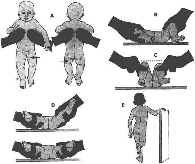
Signs of developmental dysplasia of the hip. Asymmetry of gluteal and thigh folds. Limited hip abduction as seen in flexion. Apparent shortening of the femur, as indicated by the level of the knees in flexion. Ortolani (if infant is under 4 weeks of age). Positive Trendelenburg sign or gait (if child is weight bearing).

Condition Definition Age at Onset/ Sex Difference Treatment Nursing Considerations
Clubfoot Downward, inward rotation of one or both feet: talipes equinovarus (95%) Newborn (congenital); twice as common in Series of casts changed weekly followed by Denis Browne splint and then corrective shoes (severe cases-surgery) Care of child in cast/brace Stress need for follow-up Encourage compliance
Developmental hip dysplasia Abnormal development of hip joint (most frequently unilateral) Newborn (congenital); more common in girls Newborn-Pavlik harness; older infant or toddler- possible surgery, spica cast Early identification Care of child in traction/cast: check circulation; turn q2h while cast
Osteomyelitis Most frequently occurring bone infection among children 5-14 years; twice as common in boys Blood cultures to diagnose causative organisms-select appropriate antibiotic; bed rest, immobilization with splint or cast Encourage compliance Check for other anomalies (e.g., spina bifida) Care of child in splint/cast Provide diversion Pain medications/antibiotics per MD order
Legg-CalvéPerthes Aseptic necrosis of the head of the femur (cause unknown) Peak: 4-8 years; range: 3-12 years; 5 times more common in boys; 10 times more common in whites than nonwhites Conservative therapy lasts 2-4 years, usually begins with bed rest and traction, followed by non-weight-bearing devices such as brace, cast Early identification Care of child in traction/ cast: check for frayed pulley ropes Provide diversion Assist child and family to cope with child’s prolonged immobility
Juvenile rheumatoid arthritis Chronic systemic inflammatory disease (cause unknown) Peak: 2-5 years and 9-12 years; more common in girls Prevent joint deformity by exercise, splints, medications (steroids, ASA); relieve symptoms (as per adult with arthritis) Care of child in brace/splint Provide diversion Encourage compliance
Scoliosis Lateral curvature of the spine (cause unknown) Adolescence; more common in girls Milwaukee brace; halopelvic traction; Harrington rod Care of child in traction/ cast/brace Teach that brace is worn 16-23 hours/day, 7 days/week for 6 months-2 years
Osteosarcoma Most frequently occurring bone cancer among children Adolescence (10-25 years); more common in boys and men Amputation-»prosthesis; chemotherapy Encourage compliance Promote positive self-image Prepare child for loss of limb Help cope with prosthesis, life-threatening illness Assist with grieving process

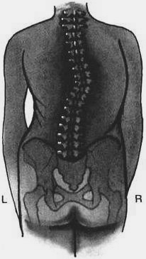
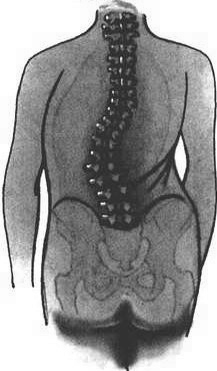
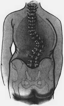
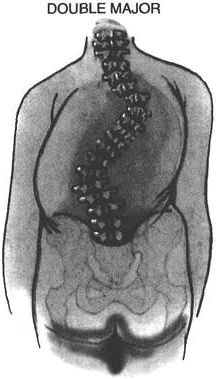
The four major curve patterns in idiopathic scoliosis.

THORACIC

90% occur on the left side at T-11

LUMBAR

70% occur on the left side at L-1 or lower

THORACOLUMBAR

80% occur on the right side at T-11 - T-12

DOUBLE MAJOR

Usually involves right-sided and lef-sided lumbar curves

Curve location is determined by the level of the apical vertebra