Cardiovascular Disorders

Congenital Heart Disease (CHD)

  1. Introduction: There are more than 35 documented types of congenital heart defects, which occur in 4 to 10 per 1000 live births. For the purpose of this review, only 5 major defects are given. Note: The content has been synthesized for ease in review and recall; for additional study aids, also contains information on congestive heart failure, and covers the most commonly used drugs, including digoxin and furosemide (Lasix).

  2. Assessment:
    1. Exact cause unknown, but related factors include:
      1. Familial history of CHD, especially in siblings, parents.
      2. Presence of other genetic defects in infant, e.g., Down syndrome, trisomy 13 or 18.
      3. History of maternal prenatal infection with rubella, cytomegalovirus, etc.
      4. High-risk maternal factors:
        1. Age: under 18 years, over 40 years.
        2. Weight: under 100 lb, over 200 lb.
        3. Maternal insulin-dependent diabetes.
      5. Maternal history of drinking during pregnancy, with resultant “fetal alcohol syndrome.”
      6. Extracardiac defects including tracheoesophageal fistula, renal agenesis, and diaphragmatic hernia.
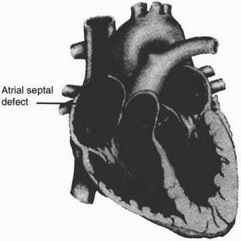
Atrial septal defect. An opening between the two atria, allowing oxygenated blood and unoxygenated blood to mix. Left-to-right shunting of blood occurs due to the higher pressure on the left side of the heart.
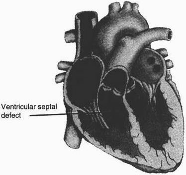
Ventricular septal defect. An opening between the two ventricles, allowing oxygenated and unoxygenated blood to mix. Shunting of blood occurs due to the higher pressure on the left side of the heart. Ventricular septal defects are classified as membranous or muscular according to location in the septum.
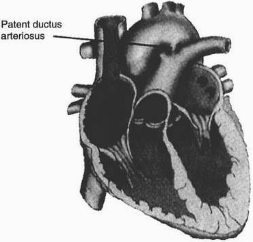
Patent ductus arteriosus. An artery that connects the aorta and the pulmonary artery during fetal life. It generally closes spontaneously within a few hours to several days after birth. Allows abnormal blood flow from the highpressure aorta to the low-pressure pulmonary artery, resulting in a left-to-right shunt.


    1. Most frequent parental complaint: difficulty feeding.
      1. Infant must be awakened to feed.
      2. Has weak suck.
      3. May turn blue when eating, especially with cyanotic defects.
      4. Infant takes overly long time to feed.
      5. Falls asleep during feeding, without finishing.
    2. Nursing observations
      1. Most frequent symptom - tachycardia, as body attempts to compensate for lack of oxygen (hypoxia), i.e., heart rate over 160 beats/minute.
      2. Tachypnea, corresponding to heart rate, i.e., respirations over 60 breaths/minute.
      3. Cyanosis due to hypoxia:
        1. Not with acyanotic defects (unless CHF is present).
        2. Always with cyanotic defects (“blue babies”).
      4. Failure to grow at a normal rate, i.e., slow weight gain, height and weight below the norm due to difficulty feeding and hypoxia.
      5. Developmental delays related to weakened physical condition.
      6. Frequent respiratory infections associated with increased pulmonary blood flow or aspiration.
      7. Dyspnea on exertion due to hypoxia, shunting of blood.
      8. Murmurs may or may not be present, e.g., patent ductus arteriosus (PDA) machinery murmur.
      9. Changes in blood pressure, e.g., coarctation - increased blood pressure in arms; decreased blood pressure in legs.
      10. Possible congestive heart failure - Note: Infants may not demonstrate distended neck veins.
      11. Cyanotic heart defects:
        1. “Tet. spells” - choking spells with paroxysmal dyspnea: severe hypoxia, deepening cyanosis; relieved by squatting, or placing infant in knee-chest position, which alters cardiopulmonary dynamics, thus increasing the flow of blood to the lungs.
        2. Clubbing of fingers and toes - due to chronic hypoxia.
        3. Polycythemia (increased red blood cells [RBCs]) with possible thrombi/emboli formation.
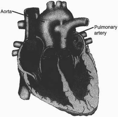
Transposition of the great arteries. The aorta and the pulmonary artery are reversed; i.e., the aorta rises from the right instead of the left ventricle, and the pulmonary artery arises from the left instead of the right ventricle. Systemic venous blood (unoxygenated blood) returns to the right side of the heart to pass through the aorta back to the body without being oxygenated because of bypassing the lungs. The pulmonary venous blood (oxygenated blood) enters the left side of the heart, returning to the lungs via the pulmonary artery. The systemic (unoxygenated) and pulmonary (oxygenated) circulations are totally separate. There must be some opening (i.e., patent ductus arteriosus or septal defect) to allow blood to mix.


Comparison of Acyanotic and Cyanotic Heart Disease
Feature Acyanotic Cyanotic
Shunting of blood

Cyanosis

Surgery Prognosis

Major types
L—> R

Not usual (unless congestive heart failure)

Usually done in one stage—technically simple

Very good/excellent
  1. Atrial septal defect (ASD)
  2. Ventricular septal defect (VSD)
  3. Patent ductus arteriosus (PDA)
  4. Coarctation of the aorta
R—>L

Always; “blue babies”

Usually done in several stages—technically complex

Guarded

  1. Tetralogy of Fallot
  2. Transposition of the great vessels


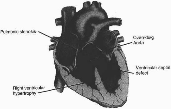
Tetralogy of Fallot. Four cardiac anomalies make up tetralogy of Fallot: a ventricular septal defect, pulmonary stenosis, an overriding aorta, and right ventricular hypertrophy. There are other associated defects in certain cases. Pulmonary stenosis results in reduction of pulmonary blood flow; the ventricular septal defect allows mixing of oxygenated and unoxygenated blood.
  1. Analysis/nursing diagnosis:
    1. Ineffective breathing pattern related to tachypnea and respiratory infection.
    2. Activity intolerance related to tachycardia and hypoxia.
    3. Altered nutrition, less than body requirements, related to difficulty in feeding.
    4. Risk for infection related to poor nutritional status.
    5. Knowledge deficit related to diagnostic procedures, condition, surgicai/medical treatments, prognosis.

  2. Nursing care plan/implementation:
    1. promote adequate oxygenation.
      1. Administer oxygen per physician’s order/prn.
      2. Use loose-fitting clothing; tape diapers loosely to avoid pressure on abdominal organs, which could impinge on diaphragm and impede respiration.
      3. Position: neck slightly hyperextended to keep airway patent; place in knee-chest (squatting) position to relieve “Tet. spell” (choking spell).
      4. Suction prn to clear the airway.
      5. Administer digoxin, per physician’s order, to slow and strengthen heart’s pumping action (refer for pediatric pulse rate norms).
    2. Goal: reduce workload of heart to conserve energy.
      1. Position: infant seat, semi - Fowler’s to promote maximum expansion of the lungs.
      2. Provide pacifier to promote psychological rest.
      3. Organize nursing care to provide periods of uninterrupted rest.
      4. Adjust physical activity according to child’s condition, capabilities to conserve energy.
      5. Provide diversion, as tolerated, to meet developmental needs yet conserve energy.
      6. Avoid extremes of temperature to avoid the stress of hypothermia/hyperthermia, which will increase the body’s demand for oxygen.
      7. Administer diuretics (Lasix), per physician’s order, to eliminate excess fluids, which increase the heart’s workload.
    3. Goal: provide for adequate nutrition.
      1. Offer low - sodium formula (Lonalac) to minimize fluid retention.
      2. Discourage foods with high or added sodium to minimize fluid retention.
      3. I&O, daily/weekly weights, and monitor for rate of growth.
      4. Limit PO feedings to 20 minutes to avoid overtiring infant. Supplement PO feeding with gavage feeding (prn with physician’s order) to meet fluid and caloric needs.
      5. Encourage foods high in potassium (prevent hypokalemia) and high in iron (prevent anemia).
    4. Goal: prevent infection.
      1. Standard precautions to prevent infection.
      2. Use good hand - washing technique.
      3. Limit contact with staff/visitors (especially children) with infections.
      4. Monitor for early symptoms and signs of infection; report stat.
    5. Goal: meet teaching needs of patient, family.
      1. Explain diagnostic procedures: blood tests, x - rays, urine, ECG, echocardiogram, cardiac catheterization.
      2. Explain condition/treatment/prognosis.
      3. Review dietary restrictions, medications.
      4. Discuss how to adjust realistically to life with congenital heart disease, activity restrictions, etc.

  3. Evaluation/outcome criteria:
    1. Child’s level of oxygenation is maintained, as evidenced by pink color in nailbeds and mucous membranes (for both light - and dark - skinned children) and ease in respiratory effort.
    2. Energy is conserved, thus reducing the heart’s workload as evidenced by vital signs within normal limits.
    3. The child’s fluid and caloric requirements are met, allowing for physical growth to occur at normal or near - normal rate.
    4. The family (and child, when old enough) verbalize their understanding of the type of CHD, its treatment and prognosis.
    5. The family and child demonstrate adequate coping mechanisms to deal with CHD.