1. Acute poststreptococcal glomerulonephritis
    1. Introduction: Acute poststreptococcal glomerulonephritis (APSGN) is a bilateral inflammation of the glomeruli of the kidneys and is the most common noninfectious renal disease of childhood. It occurs most frequently in early schoolage children, with a peak age of onset of 6-7 years; it is twice as common in boys as in girls. Like rheumatic fever, acute glomerulonephritis is thought to be the result of an antigen-antibody reaction to a streptococcus infection; however, unlike rheumatic fever, it does not tend to recur, because specific immunity is conferred following the first episode of APSGN.
    2. Assessment:
      1. Typical concerns from family about urine: change in color/appearance of urine (thick, reddish brown; decreased amounts).
      2. Acute edematous phase- usually lasts 4-10 days.
        1. Lab examination of urine:
          1. Severe hematuria.
          2. Mild proteinuria.
          3. Increased specific gravity.
        2. Hypertension
          1. Headache.
          2. Potential hypertensive encephalopathy leading to seizures, increased intracranial pressure.
        3. Mild-moderate edema: chiefly periorbital; increased weight due to fluid retention.
        4. General:
          1. Abdominal pain.
          2. Malaise.
          3. Anorexia.
          4. Vomiting.
          5. Pallor.
          6. Irritability.
          7. Lethargy.
          8. Fever.
        5. Diuresis phase:
          1. Copious diuresis.
          2. Decreased body weight.
          3. Marked clinical improvement.
          4. Decrease in gross hematuria, but microscopic hematuria may persist for weeks/ months.

Comparison of Nephrosis and Acute Poststreptococcal Glomerulonephritis

Factor Nephrosis (Nephrotic Syndrome) Acute Poststreptococcal Glomerulonephritis (APSGN)
Illness type Chronic Acute
Illness course Characterized by periods of exacerbations and remissions during many years Predictable, self-limiting, typically lasting 4-10 days (acute edematous phase)
Cause Unknown Group A (3-hemolytic streptococci
Age at onset 2-4 years Early school-age children; peaks at 6-7 years
Sex More common among boys More common among boys
Major signs and symptoms:
General Syndrome with variable pathology, massive proteinuria, hypoalbuminemia, severe edema, hyperlipidemia Hematuria, hypertension
Blood pressure Normal or decreased Elevated
Edema Generalized and severe Periorbital and peripheral
Proteinuria Massive Moderate
Serum protein level Decreased (6.1-7.9 g/dL) Increased
Serum lipid level Elevated Normal
Potassium level Normal (3.5-5 mEq/L) Increased
Treatment Symptomatic—no known cure; prednisone, cyclophosphamide, furosemide Penicillin (EES), hydralazine, furosemide
Diet Decrease sodium, increase protein Decrease sodium, decrease potassium, increase protein
Fluid restrictions Seldom necessary Necessary if output is significantly reduced
Specific nursing care Treat at home if possible; good skin care; prevent infection Treat in hospital during acute phase; monitor vital signs, especially blood pressure; on discharge, stress need to restrict activity until microscopic hematuria is gone
Prognosis Fair; subject to long-term steroid treatment and social isolation related to frequent hospitalizations/confinement during relapses; 20% suffer chronic renal failure Good; stress that recurrence is rare because specific immunity is conferred


      1. Analysis/nursing diagnosis:
        1. Fluid volume excess related to decreased urine output.
        2. Pain related to fluid retention.
        3. Altered nutrition, less than body requirements, related to anorexia and vomiting.
        4. Impaired skin integrity related to immobility.
        5. Activity intolerance related to fatigue.
        6. Knowledge deficit related to disease process, treatment, and follow-up care.
      2. Nursing care plan/implementation:
        1. Goal: monitor fluid balance, observing carefully for complications.
          1. Check and record blood pressure at least every 4 hr to monitor hypertension.
          2. Monitor daily weights.
          3. J c. Urine: strict I8<O; specific gravity and dipstick for blood every void.
          4. Note edema: extent, location, progression.
          5. Adhere to fluid restrictions if ordered.
          6. Bed rest, chiefly due to hypertension: monitor for possible development of hypertensive encephalopathy (seizures, increased intracranial pressure); report any changes stat to physician.
          7. Administer medications as ordered:
            1. Antibiotics —eradicate any lingering streptococcus infection.
            2. Antihypertensives, e.g., Apresoline.
            3. Rarely use diuretics—limited value. (4) If CHF develops—may use digoxin.
            4. Refer to Unit 10 for additional information on medications.
        2. Goal: provide adequate nutrition.
          1. Diet: low sodium, low potassium —to prevent fluid retention and hyperkalemia; high protein to replace protein being lost in the urine. Refer to Unit 9 for additional information on diets.
          2. Stimulate appetite: offer small portions, attractively prepared; meals with family or other children; offer preferred foods, if possible; encourage parents to bring in special foods, e.g., culturally related preferences.
        3. Goal: provide reasonable measure of comfort.
          1. Encourage parental visiting.
          2. Provide for positional changes, give good skin care.
          3. Provide appropriate diversion, as tolerated.
        4. Goal: prevent further infection.
          1. i a. Teach how to check urine at home: dipstick for protein and blood. (Note: occult hematuria may persist for months.)
          2. Teach activity restriction: no strenuous activity until hematuria is completely resolved.
          3. Teach family how to prepare low-sodium, low-potassium diet.
          4. Arrange for follow-up care: physician, home health nurse.
          5. Stress: subsequent recurrences are rare because specific immunity is conferred.
        5. Goal: teach child and family about APSGN/ discharge planning.
          1. Teach how to check urine at home: dipstick for protein and blood. (Note: occult hematuria may persist for months.)
          2. Teach activity restriction: no strenuous activity until hematuria is completely resolved.
          3. Teach family how to prepare low-sodium, low-potassium diet.
          4. Arrange for follow-up care: physician, home health nurse.
          5. Stress: subsequent recurrences are rare because specific immunity is conferred.
      3. Evaluation/outcome criteria:
        1. No permanent renal damage occurs.
        2. Normal fluid balance is maintained/restored.
        3. Adequate nutrition is maintained.
        4. No secondary infections occur.
        5. Child/family verbalize their understanding of the disease, its treatment, and its prognosis.

    Kawasaki Disease (Mucocutaneous Lymph Node Syndrome)

    1. Introduction: Kawasaki disease (mucocutaneous lymph node syndrome) is an acute, febrile, multisystem disorder believed to be autoimmune in nature. Affecting primarily the skin and mucous membranes of the respiratory tract, lymph nodes, and heart, Kawasaki disease has a low fatality rate (<2%), although vasculitis and cardiac involvement (coronary artery changes) may result in major complications in as many as 20-25% of children with this disease. The disease is not believed to be communicable, and the exact cause remains unknown; geographic (living near fresh water) and seasonal (late winter, early spring) outbreaks do occur. Kawasaki disease occurs in both boys and girls between 1 and 14 years of age; 80% of cases occur in children under age 5 years. It may be preceded by URI or exposure to a freshly cleaned carpet. A complete and apparently spontaneous recovery occurs within 3 to 4 wk in the majority of cases. Treatment, which is primarily symptomatic, does not appear to either enhance recovery or prevent complications, although recent research indicates that life-threatening complications and longterm disability may be avoided or minimized with early treatment (i.e., gamma globulin) to reduce cardiovascular damage.
    1. Assessment:
      1. Abrupt onset with high fever (102°-106°F) lasting more than 5 days that does not remit with the administration of antibiotics and antipyretics.
      2. Conjunctivitis—bilateral, nonpurulent.
      3. Oropharyngeal manifestations:
        1. Dry, red, cracked lips.
        2. Oropharyngeal reddening and a “strawberry” tongue.
      4. Peeling (desquamation) of the palms of the hands and the soles of the feet; begins at the fingertips and the tips of the toes; as peeling progresses, hands and feet become very red, sore, and swollen.
      5. Cervical lymphadenopathy.
      6. Generalized erythematous rash on trunk and extremities, without vesicles or crusts.
      7. Irritability, anorexia.
      8. Arthralgia and arthritis.
      9. Panvasculitis of coronary arteries: formation of aneurysms and thrombi; CHF, myocarditis, pericardial effusion, arrhythmias, mitral insufficiency, myocardial infarction (MI).
      10. Laboratory tests:
        1. Elevated: ESR.
        2. Elevated: WBC count.
        3. Elevated: platelet count.
    2. Analysis/nursing diagnosis:
      1. Hyperthermia related to high, unremitting fever.
      2. Altered oral mucous membrane and impaired swallowing related to oropharyngeal manifestations.
      3. Impaired skin integrity related to desquamation.
      4. Fluid volume deficit related to high fever and poor oral intake.
      5. Altered tissue perfusion (cardiovascular, potential/actual) related to vasculitis or thrombi.
      6. Knowledge deficit related to disease course, treatment, prognosis.
    3. Nursing care plan/implementation:
      1. Goal: reduce fever.
        1. Monitor rectal temperature every 2 hr or prn.
        2. Administer aspirin (not acetaminophen [Tylenol]) per physician’s order. (Note: aspirin is the drug of choice to reduce fever; also has anti-inflammatory effect and antiplatelet effect. Dose is 100 mg/kg/day in divided doses q6h. Monitor for signs of salicylate toxicity.)
        3. Tepid sponge baths or hypothermia blanket per physician’s order.
        4. Offer frequent cool fluids.
        5. Apply cool, loose-fitting clothes; use cotton bed linens only (no heavy blankets).
        6. Seizure precautions.
      2. Goal: provide comfort measures to oral cavity to ease the discomfort of swallowing.
        1. Good oral hygiene with soft sponge and diluted hydrogen peroxide.
        2. Apply petroleum jelly to lips.
        3. Bland foods in small amounts at frequent intervals.
        4. Avoid hot, spicy foods.
        5. Offer favorite foods from home or preferred foods from hospital selection.
      3. Goal: prevent infections and promote healing of skin.
        1. Monitor skin for desquamation, edema, rash.
        2. Keep skin clean, dry, well lubricated.
        3. Avoid soap to prevent drying.
        4. Gentle handling of skin to minimize discomfort.
        5. Provide sheepskin to lie on.
        6. Prevent scratching and itching—apply cotton mittens if necessary.
        7. Bed rest; elevate edematous extremities.
      4. Goal: prevent dehydration and restore normal fluid balance.
        1. Strict I&O.
        2. Monitor urine specific gravity q8h for increase (dehydration) or decrease (hydration).
        3. Monitor vital signs for fevers, tachycardia, arrhythmia.
        4. Monitor skin turgor, mucous membranes, anterior fontanel for dehydration.
        5. “Force” fluids.
        6. IV fluids per physician’s order.
      5. Goal: prevent cardiovascular complications.
        1. ECG monitor—report arrhythmias or tachycardia.
        2. Administer aspirin (see goal A) and highdose IV gamma globulin.
        3. Monitor for signs and symptoms of CHF: tachycardia, tachypnea, dyspnea, crackles, orthopnea, distended neck veins, dependent edema.
        4. Monitor circulatory status of extremities— check for possible development of thrombi.
        5. Stress need for long-term follow-up, including ECGs and echocardiograms.
    4. Evaluation/outcome criteria:
      1. Fever returns to normal.
      2. Oral cavity heals, and child is able to swallow.
      3. Skin heals, and no infection occurs.
      4. Normal fluid balance is restored.
      5. Normal cardiovascular functioning is reestablished, and no complications occur.
      6. Parents/child verbalize their understanding of Kawasaki disease.

    Bacterial Infections

    Introduction: Acute bacterial ear infection (acute otitis media) is common in young children, primarily because their eustachian tube is shorter and straighter than the adult’s; this allows for ready drainage of infected mucus from URIs directly into the middle ear. In some cases, acute otitis media precedes the onset of bacterial meningitis, an extremely serious and potentially fatal disease. Bacterial meningitis is a medical emergency, requiring early detection and prompt, aggressive therapy to prevent permanent neurological damage or death. (Refer to section on hydrocephalus, p. 274). Serous otitis (chronic) may result in hearing impairment or loss but is not likely to result in meningitis. Refer to Myringotomy, Table 5-19.


    1. Acute otitis media
      1. Assessment:
        1. Fever.
        2. Pain in affected ear. Infant may not complain of pain but may tug at ear, cry, shake head, refiise to lie down.
        3. Malaise, irritability, anorexia (possibly vomiting).
        4. May have symptoms and signs of URI: rhinorrhea, coryza, cough.
        5. Diminished response to sound.


    TABLE 5-19 Pediatric Surgery: Nursing Considerations

    Surgical Procedure Specific Nursing Care
    TONSILLECTOMY (the most frequently performed pediatric surgical procedure)
    Preoperative: check bleeding and clotting times
    Postoperative:
    • Position—place on abdomen or semiprone with head turned to side to prevent aspiration
    • Observe for most frequent complication—hemorrhage (frequent swallowing, emesis of bright red blood, shock)
    Prevent bleeding:
    • Do not suction — may cause bleeding
    • Do not encourage coughing, clearing throat, or blowing nose—may aggravate operative site and cause bleeding
    • Minimize crying
    Decrease pain:
    • Offer ice collar to decrease pain and for vasoconstriction, but do not force
    • Acetaminophen for pain (no aspirin)
    Nutrition:
    • NPO initially, then cool, clear fluids such as cool water, crushed ice, flavored icepops, dilute (noncitrus) fruit juice
    • No red or brown fluids (punch, Jell-O, icepops, colas), citrus juices, warm fluids (tea, broth), toast, milk/ice cream/pudding, carbonated sodas; progress to soft, bland foods; milk products can increase production of mucus
    Teach parents/discharge planning:
    • Signs and symptoms of infection; call physician promptly
    • 5-10 days postoperatively, expect slight bleeding
    • Continue soft, bland diet as tolerated
    MYRINGOTOMY (“tubes”)
    Postoperative:
    • Position—place with operated ear down, to allow for drainage. Expect moderate amount of purulent drainage initially
    • Keep external ear canal clean and dry
    Teach parents/discharge planning:
    • Need to keep water out of ear—use special earplugs when bathing or swimming
    • "Tubes" will remain in place 3-7 mo and then fall out spontaneously (with healing of eardrum)
    (Observe same principles of preoperative and postoperative care as for adult GI surgery)
    NPO until bowel sounds return (24—48 hr)
    APPENDECTOMY
    If appendix ruptured preoperatively or intraoperatively, position in semi-Fowler’s and implement wound precautions; administer antibiotics as ordered
    Monitor for signs and symptoms of peritonitis
    Typical course: speedy recovery, with discharge in about 2-3 days and excellent prognosis
    HERNIORRHAPHY (umbilical/inguinal)
    Umbilical: increased incidence in black infants
    Inguinal: increased incidence in boys
    Preoperative: monitor for possible complication of strangulation
    Routine postoperative GI surgery care
    Prognosis—excellent, with discharge 24-48 hr postoperatively
      1. Analysis/nursing diagnosis:
        1. Pain related to pressure of pus/purulent material on eardrum.
        2. Risk for injury/infection related to complication of meningitis.
      2. Nursing care plan/implementation:
        1. Goal: eradicate infection and prevent further complications (meningitis). Administer antibiotics as ordered.
        2. Goal: relieve pain and promote comfort.
          1. Administer decongestants as ordered.
          2. Offer analgesics/antipyretics to provide symptomatic relief and to decrease fever.
        3. Goal: health teaching.
          1. Teach parents that the child needs to finish all medication, even though child will seem clinically better within 24-48 hr.
          2. Review appropriate measures to control fever: antipyretics, cool sponges.
      3. Evaluation/outcome criteria:
        1. Infection is eradicated, no complications.
        2. Child appears to be comfortable.
    1. Bacterial meningitis
      1. Assessment:
        1. Abrupt onset: initial sign may be a seizure, following an episode of URI/acute otitis media.
        2. Chills and fever.
        3. Vomiting; may complain of headache, neck pain.
        4. Photophobia.
        5. Alterations in level of consciousness: delirium, stupor, increased intracranial pressure.
        6. Nuchal rigidity.
        7. Opisthotonos position: head is drawn backward into overextension.
        8. Hyperactive reflexes related to CNS irritability.
      2. Analysis/nursing diagnosis:
        1. Risk for infection related to communicability of meningitis.
        2. Risk for injury related to CNS irritability and seizures.
        3. Pain related to nuchal rigidity, opisthotonos position, increased muscle tension.
        4. Sensory/perceptual alterations related to seizures and changes in level of consciousness.
        5. Altered nutrition, less than body requirements, related to fever and poor oral intake.
        6. Knowledge deficit regarding diagnostic procedures, condition, treatment, prognosis.
      3. Nursing care plan/implementation:
        1. Goal: prevent spread of infection.
          1. Institute standard precautions.
          2. Enforce strict hand washing.
          3. Institute and maintain respiratory isolation for minimum of 24 hr after starting IV antibiotics, at which time child is no longer considered to be communicable and can be removed from isolation.
          4. Supervise all visitors in isolation techniques.
          5. Identify family members and others at high risk: do cultures (Haemophilus influenzae, Escherichia colt, etc.); possibly begin prophylactic antibiotics, e.g„ rifampin.
          6. Treat with IV antibiotics (as ordered) as soon as possible after admission (after cultures are obtained); continue 10-14 days (until cerebrospinal fluid [CSF] culture is negative and child appears clinically improved).
          7. Anticipate large-dose IV medications only—administer slowly in dilute form to prevent phlebitis.
          8. Restrain as needed to maintain IV.
        2. Goal: promote safety and prevent injury/ seizures.
          1. Maintain seizure precautions. Give anticonvulsants, as ordered (e.g., phenytoin).
          2. Place child near nurses’ station for maximum observation; provide private room for isolation.
          3. Minimize stimuli: quiet, calm environment.
          4. Restrict visitors to immediate family.
          5. Position: HOB slightly elevated to decrease intracranial pressure. (If opisthotonos: side-lying, for comfort and safety.)
        3. Goal: maintain adequate nutrition.
          1. NPO or clear liquids initially; supplement with IVs, because child may be unable to coordinate sucking and swallowing.
          2. Offer diet for age, as tolerated—child may experience anorexia (due to disease) or vomiting (due to increased intracranial pressure).
          3. Monitor I&O, daily weights.
      4. Evaluation/outcome criteria:
        1. No spread of infection noted; immunize all children against influenzae type B
        2. Safety maintained.
        3. Adequate nutrition and fluid intake maintained.
        4. Child recovers without permanent neurological damage (e.g., seizure disorders, hydrocephalus).
    2. Infestations
      1. Lice (pediculosis)
        1. Introduction: In children, the most common form of lice is pediculosis capitis, or head lice. This parasite feeds on the scalp, and its saliva causes severe itching. Head lice are frequently associated with the sharing of combs and brushes, hats, and clothing; thus, they are more common in girls, especially those with long hair. Lice are also associated with overcrowded conditions and poor hair hygiene.
        1. Assessment:
          1. Severe itching of scalp.
          2. Visible eggs/nits on shafts of hair.
        2. Analysis/nursing diagnosis:
          1. Impaired skin integrity related to infestation of scalp with lice.
          2. Risk for impaired skin integrity related to severe pruritus of scalp.
          3. Knowledge deficit related to transmission and prevention of disease and treatment regimen.
        3. Nursing care plan/implementation:
          1. Goal: eradicate lice infestation. Apply permethrin as drug of choice for infants and children—rub shampoo in for 4-5 min, then comb with fine-tooth comb to remove dead lice and nits (eggs).
          2. Goal: prevent spread of lice.
            1. Wear gloves and cap to protect self.
            2. Inspect other family members; treat pm with pediculocide.
            3. Wash all clothes and linens to kill any lice that may have fallen off the child’s hair.
            4. Encourage short hair, if acceptable.
            5. Teach preventive measures: do not share comb, brushes, hats.
        4. Evaiuation/outcome criteria: lice are eradicated and do not spread.
      1. Pinworms (enterobiasis)
        1. Introduction: In children, the most common helminthic infestation is pinworms. Infestation usually occurs when the child places fingers (and the pinworm eggs) into the mouth. Breaking the anus-to-niouth contamination cycle can best be accomplished by good hygiene, especially hand washing before eating and after toileting. If one family member has pinworms, it is highly likely that other family members are also infested; therefore, treat the entire family to eradicate the parasite. Pinworms are easily eradicated with antiparasitic medications.
        2. Assessment:
          1. Intense perianal itching.
          2. Visible pinworms in the stool,
          3. Vague abdominal discomfort.
          4. Anorexia and weight loss.
        3. Analysis/nursing diagnosis:
          1. Risk for infection/injury related to the anus-to-mouth contamination cycle of pinworm infestation, severe rectal itching.
          2. Knowledge deficit related to transmission and prevention of disease and treatment regimen.
        4. Nursing care plan/implementation:
          1. Goal: eradicate pinworm infestation. Treat all family members simultaneously with an antiparasitic agent, e.g., Vermox, Povan.
          2. Goal: prevent spread of pinworms.
            1. Launder all underwear, bed linens, and towels in hot soapy water to kill eggs.
            2. Teach family members the importance of good hygiene, especially hand washing before eating (or preparing food) and after toileting. Stress to children to keep their fingers out of their mouths.
        5. Evaiuation/outcome criteria: Pinworms are eradicated and do not spread; reinfestation does not occur.

    Accidents: Ingestions and Poisonings

    1. General principles of treatment for ingestions and poisonings:
      1. Prevention: refer to section on toddler safety, p. 214.
      2. How to induce vomiting:
        1. Drug of choice—syrup of ipecac (available over the counter; does not require a physician’s order). Families with young children should keep this medication on hand in case of accidental poisoning.
        2. Dose:
          1. 30 mL for adolescents over 12 yr; repeat dosage once if vomiting has not occurred within 20 min.
          2. 15 mL for children 1-12 yr; repeat dosage once if vomiting has not occurred within 20 min. Note: Do not administer to infants less than I yr of age without physician’s order.
          3. 10 mL for infants 6-12 mo; do not repeat dosage.
        3. Follow dose of ipecac with 4-8 oz of tap water or as much water as child will drink. In young children, give water first because child may refuse to drink anything else after tasting the ipecac.
        4. The child must vomit the syrup of ipecac to avoid its being absorbed and causing potentially fatal cardiotoxicity (cardiac arrhythmias, atrial fibrillation, severe heart block). If child does not vomit within 20 min of seconddose, manually stimulate gag reflex (use spoon to touch back of throat) or assist with gastric lavage.
      3. When not to induce vomiting:
        1. Child is stuporous or comatose.
        2. Poison ingested is a corrosive substance or petroleum distillate.
        3. Child is having seizures.
        4. Child is in severe shock.
        5. Child has lost the gag reflex.
    2. Salicylate poisoning
      1. Assessment:
        1. Determine how much aspirin was ingested, when, which type.
        1. Evaluate salicylate levels: normal, 0; therapeu-l tic range = 15-30 mg/dL; toxic, >30 mg/dL.|
        2. Early identification of mild toxicity:
          1. Tinnitus (ringing in the ears),
          2. Changes in vision, dizziness.
          3. Sweating.
          4. Nausea, vomiting, abdominal pain.
        3. Immediate recognition of salicylate poisoning:
          1. Hyperventilation (earliest sign).
          2. Fever—may be extremely high (105°106°F).
          3. Respiratory alkalosis or metabolic acidosis.
          4. Late signs: bleeding tendencies, severe electrolyte disturbances, liver or kidney failure.
      1. Analysis/nursing diagnosis:
        1. Ineffective breathing patterns related to hyperventilation/ respiratory alkalosis.
        2. Fluid volume deficit related to increased insensible loss of fluids through hyperventilation, increased loss of fluids through vomiting, and increased need for fluids due to hyperpyrexia (fever).
        3. Risk for injury related to bleeding.
        4. Anxiety related to parental/child feelings of guilt, uncertainty as to outcome, invasive nature of treatments.
        5. Knowledge deficit regarding accident prevention.
      2. Nursing care plan/implementation:
        1. Goal: promote excretion of salicylates.
          1. If possible, induce vomiting using syrup of ipecac (save, bring to emergency room).
          2. Assist with gastric lavage, if appropriate.
          3. Administer activated charcoal as early as possible.
          4. Assist with hemodialysis, as ordered, to promote excretion of salicylates and fluids.
          5. Administer IV fluids, as ordered.
        2. Goal: restore fluid and electrolyte balance.
          1. Monitor I&O, urinalysis, specific gravity.
          2. Prepare sodium bicarbonate, administer as ordered to correct metabolic acidosis.
          3. Monitor IV fluids and electrolytes.
          4. NPO initially (nasogastric [NG] tube).
        3. Goal: reduce temperature.
          1. No aspirin or acetaminophen, which might further complicate bleeding tendencies or lead to liver or kidney damage.
          2. Supportive measures: cool soaks, ice packs to armpits/groin, hypothermia blanket.
        4. Goal: prevent bleeding and possible hemorrhage.
          1. Monitor urine and stools for occult blood.
          2. Insert NG tube to detect gastric bleeding,
          3. Observe for petechiae, bruising; monitor laboratory values for Hct and Hgb.
          4. Administer vitamin K as ordered to correct bleeding tendencies.
        5. Goal: health education to prevent another accidental poisoning:
          1. Teach principles of poison prevention.
          2. Stress need to avoid accidental overdose with over-the-counter medications or dosage mix-ups.
          3. Allow child/parents to verbalize guilt, but avoid blaming or scapegoating.
      3. Evaluation/outcome criteria:
        1. Aspirin is successfully removed from child’s body without permanent damage.
        2. Fluid and electrolyte balance is restored and maintained.
        3. Child is afebrile.
        4. Bleeding is controlled, no hemorrhage occurs.
        5. No further episodes of poisoning occur.
    1. Acetaminophen poisoning
      1. Assessment:
        1. Determine how much acetaminophen was ingested, when, and which type.
        2. Evaluate acetaminophen levels: normal = 0; therapeutic range = 15-30 p,g/mL; toxic = 150 jig/mL 4 hr after ingestion.
        3. Initial period (2-4 hr after ingestion): malaise, nausea, vomiting, anorexia, diaphoresis, pallor.
        4. Latent period (1-3 days after ingestion): clinical improvement with asymptomatic rise in liver enzymes.
        5. Hepatic involvement (may last 7 days or may be permanent): pain in right upper quadrant (RUQ), jaundice, confusion, hepatic encephalopathy, clotting abnormalities.
        6. Gradual recuperation.
      2. Analysis/nursing diagnosis:
        1. Altered tissue perfusion related to hepatic necrosis.
        2. Fluid volume deficit related to increased loss of fluids secondary to vomiting and diaphoresis.
        3. Risk for injury related to bleeding and clotting disorders.
        4. Anxiety related to parental/child feelings of guilt, uncertainty as to outcome, and invasive nature of treatments.
        5. Knowledge deficit regarding accident prevention.
      3. Nursing care plan/implementation:
        1. Goal: promote excretion of acetaminophen.
          1. If possible, induce vomiting; save, bring to emergency department.
          2. Assist with gastric lavage, if appropriate.
          3. Administer activated charcoal.
          4. Assist with obtaining acetaminophen level 4 hr after ingestion.
        2. Goal: prevent permanent liver damage.
          1. Treatment must begin as soon as possible; therapy begun later than 10 hr after ingestion has no value.
          2. Administer the antidote (acetylcysteine \Mucomyst]) per physicians order. Usually administered through NG tube because of offensive odor. Given as one loading dose and 17 maintenance doses.
          3. Monitor hepatic functioning—assist with obtaining specimens and check results frequently; be aware that liver enzymes will rise and peak within 3 days and then should rapidly return to normal.
        1. Goal: restore fluid and electrolyte balance.
          1. Monitor vital signs and perform neurological checks every 2-4 hr and prn.
          2. Monitor I&O; urine analysis, including specific gravity; and weight.
          3. Monitor IV fluids as ordered.
        2. Goal: prevent bleeding.
          1. Assist in monitoring child’s PT; notify physician of significant changes.
          2. Monitor urine and stool for occult blood,
          3. Observe for and report any petechiae or unusual bruising.
        3. Goal: health education to prevent another accidental poisoning. (See goal 5, Nursing care plan/implementation, Salicylate poisoning.)
      1. Evaluation/outcome criteria:
        1. Acetaminophen is successfully removed from child’s body.
        2. Normal liver functioning is reestablished.
        3. Fluid and electrolyte balance is restored and maintained.
        4. No further episodes of poisoning occur.
    1. Lead poisoning
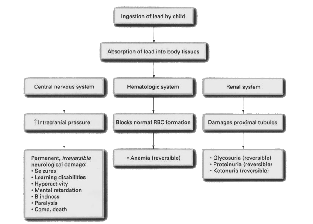
      1. Introduction: Lead poisoning is a heavy-metal poisoning that occurs from ingestion or inhalation of lead. In children, this is most common in the toddler age group (1-3 yr) and is usually a chronic type of poisoning that occurs as the result of repeated ingestions of lead. Children who engage in the practice of pica, the ingestion of nonnutritive substances, often ingest lead in flecks of lead-based paint from walls, furniture, or toys. In addition, research demonstrates that the parent-child relationship is a significant variable in lead poisoning; typically, there is a lack of adequate parental supervision that enables the child to engage in pica repeatedly over a fairly long time, until symptoms of lead poisoning become evident. (Figure 5-13 shows the pathophysiological effects of lead poisoning.)

        FIGURE 5-13 Pathophysiological effects of lead poisoning.

      2. Assessment:
        1. Investigate history of pica (ingestion of nonnutritive substance).
        2. Evaluate parent-child relationship.
        3. Chronic lead poisoning: vague, crampy abdominal pain; constipation; anorexia and vomiting; listlessness.
        4. Neurological, renal, hematological effects: see Figure 5-13.
        5. “Blood-lead line”—bluish black line seen in gums.
        6. X-rays: lead lines in long bones and flecks of lead in GI tract.
        7. Elevated serum-blood-lead levels: ^20 pg/dL requires clinical management; ^45 pg/dL requires parenteral chelating therapy.
      3. Analysis/nursing diagnosis:
        1. Altered thought processes related to neurotoxicity.
        2. Activity intolerance (and risk for infection) related to anemia.
        3. Altered urinary elimination related to excretion of lead by kidneys.
        4. Pain related to lead poisoning and its treatment.
        5. Knowledge deficit related to etiology of lead poisoning.
      4. Nursing care plan/implementation:
        1. Goal: promote excretion of lead.
          1. Administer chelating agents (EDTA [IM or IV], BAL (IM only]) as ordered. Chelation therapy typically continues over several days, with multiple treatments daily.
          2. Monitor kidney function carefully: the treatment itself is potentially nephrotoxic. Maintain adequate oral intake of fluids.
          3. Institute seizure precautions.
        2. Goal: prevent reingestion of lead.
          1. Determine primary source of poisoning.
          2. Eliminate source from child’s environment before discharge.
          3. Follow up with home care referral.
            1. Screen other siblings prn.
            2. Monitor “blood-lead level” of all children in the home.
        3. Goal: assist child to cope with multiple painful injections when treated with IM chelation therapy.
          1. Prepare child for treatment regimen.
          2. Stress that this is not a punishment.
          3. Rotate sites as much as possible.
          4. May use a local anesthetic, e.g., procaine, injected simultaneously with chelating agent to decrease pain of injections.
          5. Apply warm soaks to injection sites: may help lessen pain.
          6. Encourage child to self-limit gross muscle activity (which increases pain).
          7. Offer child safe outlets for anger, fear, frustration—punching bag, pounding board, clay.
          8. Offer opportunity for medical play with empty syringes, etc.
        4. Goal: health teaching.
          1. Stress (to child and parents) that removing the lead is the only way to prevent permanent, irreversible neurological damage (irreversible damage may have already occurred).
          2. Teach that the chelating agent binds with the lead and promotes its excretion through the kidneys.
      5. Evaluation/outcome criteria:
        1. Lead is successfully removed from child’s body without permanent damage.
        2. No further episodes of lead poisoning.
        3. Child copes successfully with the disease and its treatment.