DISORDERS AFFECTING PROTECTIVE FUNCTIONS
Immunity and Communicable Diseases
-
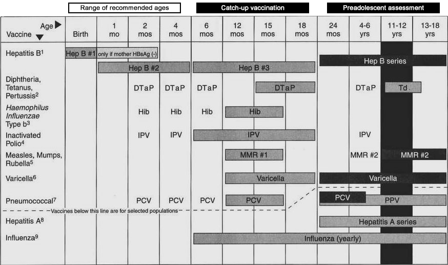
Recommended schedule for active immunization of healthy infants and children.
-
Assessment: common side effects of immunizations (occur 24-48 hours after immunization except as noted):
-
Soreness, redness, tenderness, or lump at injection site.
-
Fever: brief, mild to moderate.
-
Crankiness and fussiness; anorexia; drowsiness.
-
Measles-coryza and rash 7-10 days after immunization.
-
Rubella-arthralgia and arthritis-like symptoms 2 weeks after immunization.
-
Nursing interventions/home care for infant or child who has been immunized:
-
Explain to parents the reason for each immunization and common side effects.
-
Suggest acetaminophen for fever or discomfort.
-
Extra affection and closeness-cuddling, soothing, rocking.
-
Teach parents to notify MD of untoward, serious, or prolonged side effects.
-
Contraindications/precautions to immunizations:
-
Child who has a severe febrile illness (upper respiratory infection [URI], gastroenteritis, or any fever).
-
Child with alteration in skin integrity: rash, eczema.
-
Child with alteration in immune system; steroids; chemotherapy, radiation therapy; human immunodeficiency virus (HIV)/acquired immunodeficiency syndrome (AIDS) (no live virus vaccine).
-
Child with a known allergic reaction to previous immunization or substance in the immunization.
-
Recent recipient of blood/blood products.
-
Childhood communicable diseases. Basic principles of care:
-
Standard precautions to prevent communicability/infection.
-
Fever control with acetaminophen.
-
Extra fluids for hydration.
-
General home care procedures: comfort measures/ supportive care.
TABLE 5-16. Recommended Childhood Immunization Schedule United States, 2002
This schedule indicates the recommended ages for routine administration of currently licensed childhood vaccination as December 1, 2001, for children through age 18 years. Any does not given at the recommended age should be given at any subsequent visit when indicated and feasible. indicates age group that warrant special effort to administer those vaccines not previously given. Additional vaccines may be licensed and recommended during the year. Licensed combination vaccines may be used whenever any components of the combination are indicated and the vaccine's other components are not contraindicated. Providers should consult the manufacturers' package insertss for detailed recommendations.
Reye Syndrome
-
Introduction: Reye syndrome, first described as a disease entity in the mid-1960s, is a multisystem disorder primarily affecting children between 6 and 12 years of age. Although not truly a “communicable disease,” studies have confirmed a relationship between aspirin administration during a viral illness (chickenpox, flu) and the onset of Reye syndrome. The exact cause remains unknown. Reye syndrome is characterized by acute metabolic encephalopathy and fatty degeneration of the visceral organs, particularly the liver. Earlier diagnosis, more sophisticated monitoring equipment, and more aggressive treatment have greatly improved the survival rate of children with Reye syndrome; recovery is generally rapid in those children who do survive.
-
Assessment:
-
Onset typically follows a viral illness, just as child appears to be recovering.
-
Early signs and symptoms:
-
Rapidly progressing behavioral changes: irritability, agitation, combativeness, hostility, confusion, apathy, lethargy.
-
Vomiting, which becomes progressively worse.
-
Rapidly progressive neurological deterioration:
-
Cerebral edema and increased intracranial pressure.
-
Alteration in level of consciousness from lethargy through coma, decerebrate posturing, and respiratory arrest.
-
Liver dysfunction, necrosis, and failure:
-
Elevated serum alanine aminotransferase (ALT) (serum glutamic-oxaloacetic transaminase [SGOT]), aspartate aminotransferase (AST) (serum glutamate pyruvate transaminase [SGPT]), lactate dehydrogenase (LDH), serum-ammonia levels.
-
Severe hypoglycemia.
-
Increased prothrombin time, coagulation defects, and bleeding.
-
Analysis/nursing diagnosis:
-
Altered cerebral tissue perfusion related to cerebral edema and increased intracranial pressure.
-
Altered hepatic tissue perfusion related to fatty degeneration of the liver.
-
Risk for injury related to coagulation defects and bleeding.
-
Knowledge deficit related to diagnosis, course of disease, treatment, and prognosis.
-
Nursing care plan/implementation:
-
Goal: reduce intracranial pressure.
-
Child is admitted to pediatric intensive care unit (PICU) for intensive nursing care, continuous observation, and monitoring.
-
Monitor neurological status and vital signs continuously.
-
Assist with/prepare for numerous invasive procedures, including endotracheal (ET) tube/mechanical ventilation and intracranial pressure monitor.
-
Monitor closely for the development of seizures; institute seizure precautions.
-
Position: elevate head of bed (HOB) 30-45 degrees.
-
Administer medications as ordered:
-
Osmotic diuretics (mannitol) to decrease I CP.
-
Diuretics (Lasix) to decrease CSF production.
-
Anticonvulsants (Dilantin, phenobarbital).
-
Goal: restore and maintain fluid and electrolyte balance, including perfusion of liver.
-
Administer IV fluids per physician’s order-usually 10% glucose (or higher).
-
Strict I&O.
-
Prepare for/assist with Foley catheter placement, CVP, ICP monitor, NG tube, etc.
-
Monitor serum electrolyte lab values.
-
Goal: prevent injury and possible bleeding.
-
Observe child for petechiae, unusual bruising, oozing from body orifices or tubes, frank hemorrhage.
-
Check all urine and stool for occult blood.
-
Monitor lab values, including prothrombin time (PT), partial thromboplastin time (PTT), platelets.
-
Administer blood products per physician’s order.
-
Goal: provide parents with thorough understanding of Reye syndrome.
-
Primary nurse assigned to provide care and follow through with teaching.
-
Encourage parents’ presence, even in PICU-explain all equipment and procedures in simple, direct terms.
-
Provide factual, honest, and complete information regarding disease, diagnosis, and prognosis.
Autoimmune Disorders
Streptococcus Infections/Sequelae
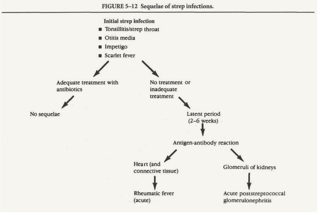
Introduction: Group A beta-hemolytic streptococcus is a common infectious organism that causes illness in children and is highly contagious. In themselves, the diseases caused by streptococcus do not seem very serious: strep throat, otitis media, impetigo, or scarlet fever. The most common treatment for strep is a full course of antibiotic therapy: 10 days of penicillin (or, if allergic, erythromycin). With adequate therapy, generally no sequelae are seen. If the strep is not treated, or is only partially treated, the sequelae include serious systemic diseases, with potentially long-term effects. If the effect is manifested primarily in the
heart (carditis), it is acute rheumatic fever. If the effect is manifested primarily in the kidneys, it is
acute glomerulonephritis
- Rheumatic fever
-
Introduction:Introduction: Rheumatic fever is an acute, systemic, inflammatory disease affecting multiple organs and systems: heart, joints, CNS, collagenous tissue, etc. Thought to be autoimmune in nature, it most commonly follows a streptococcus infection and occurs primarily in school-age children. In addition, it tends to recur, and the risk of permanent heart damage increases with each subsequent attack of rheumatic fever.
-
Assessment:
Major manifestations (modified Jones criteria)
-
Carditis: tachycardia, cardiomegaly, murmur, congestive heart failure (CHF).
-
Migratory polyarthritis: swollen, hot, red, and excruciatingly painful large joints; migratory and reversible.
-
Sydenham chorea (St. Vitus Dance): sudden, aimless, irregular movements of the extremities; involuntary facial grimaces, speech disturbances, emotional lability, muscle weakness; completely reversible.
-
Erythema marginatum: reddish pink rash most commonly found on the trunk; nonpruritic, macular, clear center, wavy but clearly marked border; transient.
-
Subcutaneous nodules: small, round, freely movable, and painless swellings usually found over the extensor surfaces of the hands/feet or bony prominences; resolve without any permanent damage.
-
Minor manifestations
-
Clinical
-
Previous history of rheumatic fever.
-
Arthralgia.
-
Fever-normal in morning, rises in mid-afternoon, normal at night.
-
Laboratory
-
Increased erythrocyte sedimentation rate (ESR).
-
Positive C-reactive protein.
-
Leukocytosis.
-
Anemia.
-
Prolonged P-R/Q-T intervals on ECG.
-
Supportive evidence
-
Recent history of streptococcus infection:
-
Strep throat/tonsilitis.
-
Otitis media.
-
Impetigo.
-
Scarlet fever.
-
Positive throat culture for streptococcus.
-
Increased ASO titer: indicates presence of streptococcus antibodies; begins to rise in 7 days, reaches maximum level in 4-6 weeks.
-
Analysis/nursing diagnosis:
-
Decreased cardiac output related to carditis.
-
Pain related to migratory polyarthritis.
-
Risk for injury related to chorea.
-
Diversional activity deficit related to lengthy hospitalization and recuperation.
-
Knowledge deficit related to preventing cardiac damage, relieving discomfort, and preventing injury.
-
Ineffective management of therapeutic regimen with long-term antibiotic therapy and follow-up care.
-
Nursing care plan/implementation:
-
Goal: prevent cardiac damage.
-
Hospitalization, with strict bed rest.
-
Monitor apical pulse for changes in rate, rhythm, murmurs.
-
Evaluate tolerance of increased activity by apical rate: if heart rate increases by more than 20 beats/min over resting rate, child should return to bed.
-
Offer low-sodium diet to prevent fluid retention.
-
Administer oxygen, digoxin/L«six as ordered (if CHF develops).
-
Goal: relieve discomfort.
-
Use bed cradle to keep linens from resting on painful joints.
-
Administer aspirin as ordered to relieve pain.
-
Move child carefully, minimally-support joints.
-
Do not massage; do not perform ROM exercises; do not apply splints; do not apply heat/cold. All these treatments will cause increased pain and are not needed, because no permanent deformities will result from this type of arthritis.
-
Goal: promote safety and prevent injury related to chorea.
-
Use side rails: elevated, padded.
-
Restrain in bed if necessary.
-
No oral temperatures-child may bite thermometer.
-
Spoon-feed-no forks or knives, to prevent injury to oral cavity.
-
Assist with all aspects of ADL until child can care for own needs.
-
Goal: provide diversion as tolerated.
-
Encourage quiet diversional activities: hobbies, reading, puzzles.
-
Get homework, books; provide tutor as condition permits.
-
Encourage contact with peers: telephone calls, letters, cards.
-
Goal: encourage child and family to comply with long-term antibiotic therapy.
-
Begin antibiotics immediately, to eradicate any lingering streptococcus infection.
-
Prepare child/family for minimum of 5 years of IM injections of penicillin.
-
Stress need for exact schedule: every 4 weeks.
-
Enlist child’s cooperation with therapy, “hero” badge.
-
Goal: health teaching.
-
To encourage compliance with prolonged bed rest-stress that ultimate prognosis depends on amount of cardiac damage.
-
Teach necessity for long-term prophylactic therapy for 5 years initially (with lifelong follow-up), during dental work, childbirth, surgery (to prevent subacute bacterial endocarditis [SBE]). Instruct adolescents to avoid body piercing and tattooing for same rationale.
-
Teach rationale: permanent cardiac damage is more likely to occur with subsequent attacks of rheumatic fever.
-
Evaluation/outcome criteria:
-
No permanent cardiac damage occurs.
-
Child is free from discomfort or is able to tolerate discomfort.
-
Injuries are avoided.
-
Child’s need for diversional activity is met.
-
Child/family comply with long-term antibiotic therapy/prophylactic therapy.