DISORDERS AFFECTING FLUID-GAS TRANSPORT


Cardiovascular Disorders


Congenital Heart Disease (CHD)

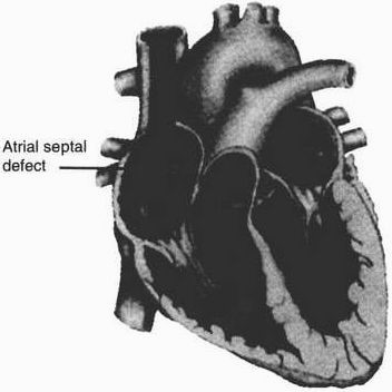
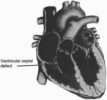
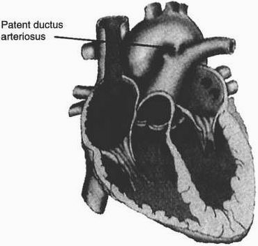
  1. Introduction: There are more than 35 documented types of congenital heart defects, which occur in 4 to 10 per 1000 live births. For the purpose of this review, only 5 major defects are given. These are presented in Figures 5-1 through 5-5. Note: The content has been synthesized for ease in review and recall; for additional study aids, the student may wish to refer to Tables 5-10 and 5-11. Unit 7 also contains information on congestive heart failure, and Unit 10 covers the most commonly used drugs, including digoxin and furosemide (Lasix).
  2. Assessment:
    1. Exact cause unknown, but related factors include:
      1. Familial history of CHD, especially in siblings, parents.
      2. Presence of other genetic defects in infant, e.g., Down syndrome, trisomy 13 or 18.
      3. History of maternal prenatal infection with rubella, cytomegalovirus, etc.
      4. High-risk maternal factors:
        1. Age: under 18 yr, over 40 yr.
        2. Weight: under 100 lb, over 200 lb.
        3. Maternal insulin-dependent diabetes.
      5. Maternal history of drinking during pregnancy, with resultant “fetal alcohol syndrome.”
      6. Extracardiac defects including tracheoesophageal fistula, renal agenesis, and diaphragmatic hernia.
FIGURE 5-1 Atrial septal defect. An opening between the two atria, allowing oxygenated blood and unoxygenated blood to mix. Left-to-right shunting of blood occurs due to the higher pressure on the left side of the heart. (From Ashwill, JW, and Droske, SC: Nursing Care of Children: Principles and Practice. WB Saunders, Philadelphia, 1997.)
FIGURE 5-2 Ventricular septal defect. An opening between the two ventricles, allowing oxygenated and unoxygenated blood to mix. Shunting of blood occurs due to the higher pressure on the left side of the heart. Ventricular septal defects are classified as membranous or muscular according to location in the septum. (From Ashwill, JW, and Droske, SC: Nursing Care of Children: Principles and Practice. WB Saunders, Philadelphia, 1997.)
FIGURE 5-3 Patent ductus arteriosus. An artery that connects the aorta and the pulmonary artery during fetal life. It generally closes spontaneously within a few hours to several days after birth. Allows abnormal blood flow from the highpressure aorta to the low-pressure pulmonary artery, resulting in a left-to-right shunt. (From Ashwill, JW, and Droske, SC: Nursing Care of Children: Principles and Practice. WB Saunders, Philadelphia, 1997.)